Analysis of CT Findings of 82 Cases of Novel Coronavirus Pneumonia in Wuhan
-
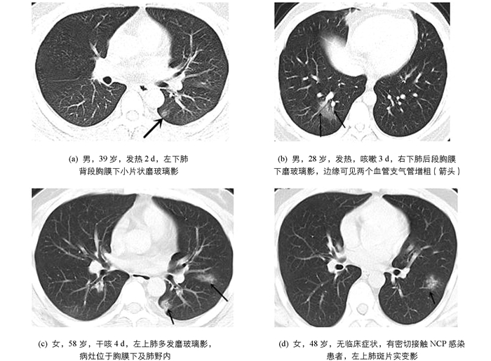
摘要:目的 分析武汉地区新型冠状病毒肺炎(novel coronavirus pneumonia,NCP)发病早期胸部CT影像表现与临床特点.方法 回顾性分析2020年1月30日-2月30日武汉科技大学附属普仁医院核酸检测确诊的82例NCP患者,所有患者均是发生临床症状(发热、咳嗽、肌肉酸痛、腹痛等)1周内或无症状初次就诊,且所有患者未经治疗,并在初次就诊时进行了胸部CT检查,由2名高年资影像诊断医师进行阅片,分析患者发病早期胸部CT影像特征,并试图发现临床各型NCP患者发病早期CT阳性病灶在各肺叶分布数量有无差异.结果 82例NCP患者中,男性38例,平均年龄(45.13±14.28)岁;女性44例,平均年龄(48.33±15.24)岁.首次胸部CT有6例NCP患者呈阴性表现,有76例胸部CT为阳性病灶.主要表现为肺内磨玻璃影(ground-glass opacity,GGO)70例(92.11%),分布于胸膜下及肺野外带58例(58/70,82.86%),同时病灶内合并血管束增粗64例(64/70,91.4%),阳性病灶累及各肺叶总数共224个,其中位于右下肺(70/224,31.25%)及左下肺叶(68/224,30.36%);斑片模糊影及其他6例(7.9%).轻型、普通型、重症及危重患者发病早期阳性病灶在各肺叶分布数量相比较差异无统计学意义(F=2.317,p > 0.05).结论 武汉地区NCP患者发病早期CT影像主要表现为多发磨玻璃影,合并血管束增粗,部位以双肺、胸膜下及肺野外带多见.对于部分患者首次胸部CT呈阴性表现,应及时近期复查.轻型、普通型、重症及危重患者发病早期阳性病灶累及肺叶的数量与后期病情轻重没有关系.Abstract:Objective To analyze the CT imaging manifestations and clinical features of early novel coronavirus pneumonia (NCP) in Wuhan area at the onset of the plague.Methods A retrospective analysis was made of 82 patients with NCP diagnosed by nucleic acid testing of Puren Hospital affiliated to Wuhan University of Science and Technology from January 30 to February 30, 2020. All the patients were asymptomatic at the first consultation or had clinical symptoms (fever, cough, muscle soreness, abdominal pain, etc.) for the first time within 1 week, and none of them had received any treatment. Then they had a chest CT examination at the first consultation. Two senior diagnostic imaging doctors reviewed the images and analyzed the CT features of the chest in the early stage of the disease and tried to find out whether there was any difference in the distribution of CT-positive lesions in each lung lobe in the early stage of NCP patients.Results Of the 82 NCP patients, 38 were males with an average age of 45.13±14.28 and 44 were females with an average age of 48.33±15.24; and 6 were negative in the first chest CT and 76 were positive. The main manifestations were ground-glass opacity (GGO) in 70 cases (70/76, 92.11%), GGO distribution in both lungs in 55 cases (55/76, 72.37%), GGO distribution under the pleura and lung field band in 58 cases (58/70, 82.86%), thickened vascular bundles in GGO lesions in 64 cases (64/70, 91.43%) and patchy shadows and others in 6 cases (6/76, 7.9%). A total of 224 positive lesions were involved in the lobes, of which 70 were located in the right lower lung (70/224, 31.25%) and 68 in the left lower lung (68/224, 30.36%); patchy shadows and 6 others (6/76, 7.9%)). There was no significant difference in the number of lung lobes involved in early-onset lesions in patients with mild, general, severe, and critical illness (F=2.317, p > 0.05).Conclusion The early CT images of patients with NCP in Wuhan showed multiple ground-glass opacities and thickened vascular bundles, which were commonly located in double lungs, subpleural and outer zone of lung fields. Some patients with negative chest CT for the first time should be reviewed in a timely manner. The number of lung lobes involved in the early stages of mild, normal, severe and critically ill patients has no relationship with the severity of the disease.
-
Key words:
- Wuhan /
- novel coronavirus pneumonia /
- CT imaging finding .
表 1 76例胸部CT阳性患者病灶分布情况分析
磨玻璃影 斑片影及其他 病灶累及肺叶总数/% 184(82.14) 40(17.86) 右上肺 18(8.04) 4(1.79) 右中肺 12(5.36) 8(3.57) 右下肺 70(31.25) 12(5.36) 左上肺 16(7.12) 6(2.68) 左下肺 68(30.36) 10(4.46) 是否双肺累及/% 双肺 55(72.37) 4(5.3) 仅单肺 15(19.74) 2(2.6) 合并血管增粗数/% 有 64(91.43) 4(66.67) 无 6(8.57) 2(33.33) 胸膜下分布数/% 有 58(82.86) 1(16.67) 无 12(17.14) 5(83.33) 表 2 轻型、普通型、重症及危重患者病灶在各肺叶的分布数量
分型 病例数 病灶肺叶数 F p 轻型 10 28 普通型 60 159 2.317 0.105 重症及危重 12 37 [1] 中华人民共和国国家卫生健康委员会.新型冠状病毒肺炎诊疗方案(第七版)[EB/OL].(2020-03-03)[2020-03-04].http://www.nhc.gov.cn/yzygj/s7653p/202003/46c9294a7dfe4cef80dc7f5912eb1989.shtml. [2] LI Q, GUAN X, WU P, et al.Early Transmission Dynamics in Wuhan, China, of Novel Coronavirus-Infected Pneumonia[J].N Engl J Med, 2020, 382(13):1199-1207. doi: 10.1056/NEJMoa2001316 [3] ROTHE C, SCHUNK M, SOTHMANN P, et al.Transmission of 2019-nCoV Infection from an Asymptomatic Contact in Germany[J].The New England Journal of Medicine, 2020, 382(10):970-971. doi: 10.1056/NEJMc2001468 [4] HUANG C L, WANG Y M, LI X W, et al.Clinical Features of Patients Infected with 2019 Novel Coronavirus in Wuhan, China[J].The Lancet, 2020, 395(10223):497-506. doi: 10.1016/S0140-6736(20)30183-5 [5] AI T, YANG Z, HOU H, et al.Correlation of Chest CT and RT-PCR Testing in Coronavirus Disease 2019(COVID-19) in China: A Report of 1014 Cases[EB/OL].(2020-02-26)[2020-03-01].https://pubs.rsna.org/doi/full/10.1148/radiol.2020200642. [6] HANSELL D M, BANKIER A A, MACMAHON H, et al.Fleischner Society:Glossary of Terms for Thoracic Imaging[J].Radiology, 2008, 246(3):697-722. doi: 10.1148/radiol.2462070712 [7] REMY-JARDIN M, REMY J, GIRAUD F, et al.Computed Tomography Assessment of Ground-Glass Opacity[J].Journal of Thoracic Imaging, 1993, 8(4):249-264. doi: 10.1097/00005382-199323000-00001 [8] WEICHERT W, WARTH A.Early Lung Cancer with Lepidic Pattern:Adenocarcinoma in Situ, Minimally Invasive Adenocarcinoma, and Lepidic Predominant Adenocarcinoma[J].Current Opinion in Pulmonary Medicine, 2014, 20(4):309-316. doi: 10.1097/MCP.0000000000000065 [9] XU Z, SHI L, WANG Y J, et al.Pathological Findings of COVID-19 Associated with Acute Respiratory Distress Syndrome[J].The Lancet Respiratory Medicine, 2020, 8(4):420-422. doi: 10.1016/S2213-2600(20)30076-X [10] HEITZMAN E R, MARKARIAN B, BERGER I, et al.The Secondary Pulmonary Lobule:a Practical Concept for Interpretation of Chest Radiographs[J].Radiology, 1969, 93(3):507-512. doi: 10.1148/93.3.507 [11] 刘发明, 丁惠玲, 龚晓明, 等.新型冠状病毒肺炎(COVID-19)的胸部CT表现与临床特点[J/OL].放射学实践, (2020-02-05)[2020-03-01].https://doi.org/10.13609/j.cnki.1000-0313.2020.03.001. [12] WANG D W, HU B, HU C, et al.Clinical Characteristics of 138 Hospitalized Patients with 2019 Novel Coronavirus-Infected Pneumonia in Wuhan, China[J].JAMA, 2020, 323(11):1061-1069. doi: 10.1001/jama.2020.1585 [13] 陈蕾, 刘辉国, 刘威, 等. 2019新型冠状病毒肺炎29例临床特征分析[J], 中华结核和呼吸杂志, 2020, 43(3):203-208. doi: 10.3760/cma.j.issn.1001-0939.2020.03.013 [14] 中华医学会放射学分会.新型冠状病毒肺炎的放射学诊断:中华医学会放射学分会专家推荐意见(第一版)[J].中华放射学杂志, 2020, 54(4):279-285. doi: 10.3760/cma.j.cn112149-20200205-00094 [15] doi: http://d.old.wanfangdata.com.cn/Periodical/syyylczz201803064 FRANQUET T.Imaging of Pulmonary Viral Pneumonia[J].Radiology, 2011, 260(1):18-39. [16] KOO H J, LIM S, CHOE J, et al.Radiographic and CT Features of Viral Pneumonia[J].RadioGraphics, 2018, 38(3):719-739. doi: 10.1148/rg.2018170048 [17] BAJANTRI B, VENKATRAM S, DIAZ-FUENTES G.Mycoplasma Pneumoniae:a Potentially Severe Infection[J].Journal of Clinical Medicine Research, 2018, 10(7):535-544. doi: 10.14740/jocmr3421w 82例武汉地区新型冠状病毒肺炎发病早期CT影像表现分析
- 收稿日期: 2020-03-18
基金项目: 江西省卫生健康委员会科技计划项目(20195254);武汉市卫生健康委员会科研基金项目(WX18D01)摘要:
目的 分析武汉地区新型冠状病毒肺炎(novel coronavirus pneumonia,NCP)发病早期胸部CT影像表现与临床特点.方法 回顾性分析2020年1月30日-2月30日武汉科技大学附属普仁医院核酸检测确诊的82例NCP患者,所有患者均是发生临床症状(发热、咳嗽、肌肉酸痛、腹痛等)1周内或无症状初次就诊,且所有患者未经治疗,并在初次就诊时进行了胸部CT检查,由2名高年资影像诊断医师进行阅片,分析患者发病早期胸部CT影像特征,并试图发现临床各型NCP患者发病早期CT阳性病灶在各肺叶分布数量有无差异.结果 82例NCP患者中,男性38例,平均年龄(45.13±14.28)岁;女性44例,平均年龄(48.33±15.24)岁.首次胸部CT有6例NCP患者呈阴性表现,有76例胸部CT为阳性病灶.主要表现为肺内磨玻璃影(ground-glass opacity,GGO)70例(92.11%),分布于胸膜下及肺野外带58例(58/70,82.86%),同时病灶内合并血管束增粗64例(64/70,91.4%),阳性病灶累及各肺叶总数共224个,其中位于右下肺(70/224,31.25%)及左下肺叶(68/224,30.36%);斑片模糊影及其他6例(7.9%).轻型、普通型、重症及危重患者发病早期阳性病灶在各肺叶分布数量相比较差异无统计学意义(F=2.317,p > 0.05).结论 武汉地区NCP患者发病早期CT影像主要表现为多发磨玻璃影,合并血管束增粗,部位以双肺、胸膜下及肺野外带多见.对于部分患者首次胸部CT呈阴性表现,应及时近期复查.轻型、普通型、重症及危重患者发病早期阳性病灶累及肺叶的数量与后期病情轻重没有关系.English Abstract
Analysis of CT Findings of 82 Cases of Novel Coronavirus Pneumonia in Wuhan
- Received Date: 2020-03-18
Abstract:
Objective To analyze the CT imaging manifestations and clinical features of early novel coronavirus pneumonia (NCP) in Wuhan area at the onset of the plague.Methods A retrospective analysis was made of 82 patients with NCP diagnosed by nucleic acid testing of Puren Hospital affiliated to Wuhan University of Science and Technology from January 30 to February 30, 2020. All the patients were asymptomatic at the first consultation or had clinical symptoms (fever, cough, muscle soreness, abdominal pain, etc.) for the first time within 1 week, and none of them had received any treatment. Then they had a chest CT examination at the first consultation. Two senior diagnostic imaging doctors reviewed the images and analyzed the CT features of the chest in the early stage of the disease and tried to find out whether there was any difference in the distribution of CT-positive lesions in each lung lobe in the early stage of NCP patients.Results Of the 82 NCP patients, 38 were males with an average age of 45.13±14.28 and 44 were females with an average age of 48.33±15.24; and 6 were negative in the first chest CT and 76 were positive. The main manifestations were ground-glass opacity (GGO) in 70 cases (70/76, 92.11%), GGO distribution in both lungs in 55 cases (55/76, 72.37%), GGO distribution under the pleura and lung field band in 58 cases (58/70, 82.86%), thickened vascular bundles in GGO lesions in 64 cases (64/70, 91.43%) and patchy shadows and others in 6 cases (6/76, 7.9%). A total of 224 positive lesions were involved in the lobes, of which 70 were located in the right lower lung (70/224, 31.25%) and 68 in the left lower lung (68/224, 30.36%); patchy shadows and 6 others (6/76, 7.9%)). There was no significant difference in the number of lung lobes involved in early-onset lesions in patients with mild, general, severe, and critical illness (F=2.317, p > 0.05).Conclusion The early CT images of patients with NCP in Wuhan showed multiple ground-glass opacities and thickened vascular bundles, which were commonly located in double lungs, subpleural and outer zone of lung fields. Some patients with negative chest CT for the first time should be reviewed in a timely manner. The number of lung lobes involved in the early stages of mild, normal, severe and critically ill patients has no relationship with the severity of the disease.- Figure 1.
- Figure 2.