Message Board

Dear readers, authors and reviewers,you can add a message on this page. We will reply to you as soon as possible!

2020 Volume 42 Issue 5
Article Contents

Zhong-xiang ZHU, Qing ZHANG, Wei XU, et al. Analysis of CT Findings of 82 Cases of Novel Coronavirus Pneumonia in Wuhan[J]. Journal of Southwest University Natural Science Edition, 2020, 42(5): 36-41. doi: 10.13718/j.cnki.xdzk.2020.05.005
Citation: Zhong-xiang ZHU, Qing ZHANG, Wei XU, et al. Analysis of CT Findings of 82 Cases of Novel Coronavirus Pneumonia in Wuhan[J]. Journal of Southwest University Natural Science Edition, 2020, 42(5): 36-41. doi: 10.13718/j.cnki.xdzk.2020.05.005

Analysis of CT Findings of 82 Cases of Novel Coronavirus Pneumonia in Wuhan

More Information
  • Corresponding author: Qing ZHANG ; 
  • Received Date: 18/03/2020
    Available Online: 20/05/2020
  • MSC: R563.1

  • Objective To analyze the CT imaging manifestations and clinical features of early novel coronavirus pneumonia (NCP) in Wuhan area at the onset of the plague. Methods A retrospective analysis was made of 82 patients with NCP diagnosed by nucleic acid testing of Puren Hospital affiliated to Wuhan University of Science and Technology from January 30 to February 30, 2020. All the patients were asymptomatic at the first consultation or had clinical symptoms (fever, cough, muscle soreness, abdominal pain, etc.) for the first time within 1 week, and none of them had received any treatment. Then they had a chest CT examination at the first consultation. Two senior diagnostic imaging doctors reviewed the images and analyzed the CT features of the chest in the early stage of the disease and tried to find out whether there was any difference in the distribution of CT-positive lesions in each lung lobe in the early stage of NCP patients. Results Of the 82 NCP patients, 38 were males with an average age of 45.13±14.28 and 44 were females with an average age of 48.33±15.24; and 6 were negative in the first chest CT and 76 were positive. The main manifestations were ground-glass opacity (GGO) in 70 cases (70/76, 92.11%), GGO distribution in both lungs in 55 cases (55/76, 72.37%), GGO distribution under the pleura and lung field band in 58 cases (58/70, 82.86%), thickened vascular bundles in GGO lesions in 64 cases (64/70, 91.43%) and patchy shadows and others in 6 cases (6/76, 7.9%). A total of 224 positive lesions were involved in the lobes, of which 70 were located in the right lower lung (70/224, 31.25%) and 68 in the left lower lung (68/224, 30.36%); patchy shadows and 6 others (6/76, 7.9%)). There was no significant difference in the number of lung lobes involved in early-onset lesions in patients with mild, general, severe, and critical illness (F=2.317, p > 0.05). Conclusion The early CT images of patients with NCP in Wuhan showed multiple ground-glass opacities and thickened vascular bundles, which were commonly located in double lungs, subpleural and outer zone of lung fields. Some patients with negative chest CT for the first time should be reviewed in a timely manner. The number of lung lobes involved in the early stages of mild, normal, severe and critically ill patients has no relationship with the severity of the disease.
  • 加载中
  • [1] 中华人民共和国国家卫生健康委员会.新型冠状病毒肺炎诊疗方案(第七版)[EB/OL].(2020-03-03)[2020-03-04].http://www.nhc.gov.cn/yzygj/s7653p/202003/46c9294a7dfe4cef80dc7f5912eb1989.shtml.

    Google Scholar

    [2] LI Q, GUAN X, WU P, et al.Early Transmission Dynamics in Wuhan, China, of Novel Coronavirus-Infected Pneumonia[J].N Engl J Med, 2020, 382(13):1199-1207. doi: 10.1056/NEJMoa2001316

    CrossRef Google Scholar

    [3] ROTHE C, SCHUNK M, SOTHMANN P, et al.Transmission of 2019-nCoV Infection from an Asymptomatic Contact in Germany[J].The New England Journal of Medicine, 2020, 382(10):970-971. doi: 10.1056/NEJMc2001468

    CrossRef Google Scholar

    [4] HUANG C L, WANG Y M, LI X W, et al.Clinical Features of Patients Infected with 2019 Novel Coronavirus in Wuhan, China[J].The Lancet, 2020, 395(10223):497-506. doi: 10.1016/S0140-6736(20)30183-5

    CrossRef Google Scholar

    [5] AI T, YANG Z, HOU H, et al.Correlation of Chest CT and RT-PCR Testing in Coronavirus Disease 2019(COVID-19) in China: A Report of 1014 Cases[EB/OL].(2020-02-26)[2020-03-01].https://pubs.rsna.org/doi/full/10.1148/radiol.2020200642.

    Google Scholar

    [6] HANSELL D M, BANKIER A A, MACMAHON H, et al.Fleischner Society:Glossary of Terms for Thoracic Imaging[J].Radiology, 2008, 246(3):697-722. doi: 10.1148/radiol.2462070712

    CrossRef Google Scholar

    [7] REMY-JARDIN M, REMY J, GIRAUD F, et al.Computed Tomography Assessment of Ground-Glass Opacity[J].Journal of Thoracic Imaging, 1993, 8(4):249-264. doi: 10.1097/00005382-199323000-00001

    CrossRef Google Scholar

    [8] WEICHERT W, WARTH A.Early Lung Cancer with Lepidic Pattern:Adenocarcinoma in Situ, Minimally Invasive Adenocarcinoma, and Lepidic Predominant Adenocarcinoma[J].Current Opinion in Pulmonary Medicine, 2014, 20(4):309-316. doi: 10.1097/MCP.0000000000000065

    CrossRef Google Scholar

    [9] XU Z, SHI L, WANG Y J, et al.Pathological Findings of COVID-19 Associated with Acute Respiratory Distress Syndrome[J].The Lancet Respiratory Medicine, 2020, 8(4):420-422. doi: 10.1016/S2213-2600(20)30076-X

    CrossRef Google Scholar

    [10] HEITZMAN E R, MARKARIAN B, BERGER I, et al.The Secondary Pulmonary Lobule:a Practical Concept for Interpretation of Chest Radiographs[J].Radiology, 1969, 93(3):507-512. doi: 10.1148/93.3.507

    CrossRef Google Scholar

    [11] 刘发明, 丁惠玲, 龚晓明, 等.新型冠状病毒肺炎(COVID-19)的胸部CT表现与临床特点[J/OL].放射学实践, (2020-02-05)[2020-03-01].https://doi.org/10.13609/j.cnki.1000-0313.2020.03.001.

    Google Scholar

    [12] WANG D W, HU B, HU C, et al.Clinical Characteristics of 138 Hospitalized Patients with 2019 Novel Coronavirus-Infected Pneumonia in Wuhan, China[J].JAMA, 2020, 323(11):1061-1069. doi: 10.1001/jama.2020.1585

    CrossRef Google Scholar

    [13] 陈蕾, 刘辉国, 刘威, 等. 2019新型冠状病毒肺炎29例临床特征分析[J], 中华结核和呼吸杂志, 2020, 43(3):203-208. doi: 10.3760/cma.j.issn.1001-0939.2020.03.013

    CrossRef Google Scholar

    [14] 中华医学会放射学分会.新型冠状病毒肺炎的放射学诊断:中华医学会放射学分会专家推荐意见(第一版)[J].中华放射学杂志, 2020, 54(4):279-285. doi: 10.3760/cma.j.cn112149-20200205-00094

    CrossRef Google Scholar

    [15] FRANQUET T.Imaging of Pulmonary Viral Pneumonia[J].Radiology, 2011, 260(1):18-39.

    Google Scholar

    [16] KOO H J, LIM S, CHOE J, et al.Radiographic and CT Features of Viral Pneumonia[J].RadioGraphics, 2018, 38(3):719-739. doi: 10.1148/rg.2018170048

    CrossRef Google Scholar

    [17] BAJANTRI B, VENKATRAM S, DIAZ-FUENTES G.Mycoplasma Pneumoniae:a Potentially Severe Infection[J].Journal of Clinical Medicine Research, 2018, 10(7):535-544. doi: 10.14740/jocmr3421w

    CrossRef Google Scholar

  • 加载中
通讯作者: 陈斌, bchen63@163.com
  • 1. 

    沈阳化工大学材料科学与工程学院 沈阳 110142

  1. 本站搜索
  2. 百度学术搜索
  3. 万方数据库搜索
  4. CNKI搜索

Figures(2)  /  Tables(2)

Article Metrics

Article views(5792) PDF downloads(129) Cited by(0)

Access History

Analysis of CT Findings of 82 Cases of Novel Coronavirus Pneumonia in Wuhan

    Corresponding author: Qing ZHANG ; 

Abstract:  Objective To analyze the CT imaging manifestations and clinical features of early novel coronavirus pneumonia (NCP) in Wuhan area at the onset of the plague. Methods A retrospective analysis was made of 82 patients with NCP diagnosed by nucleic acid testing of Puren Hospital affiliated to Wuhan University of Science and Technology from January 30 to February 30, 2020. All the patients were asymptomatic at the first consultation or had clinical symptoms (fever, cough, muscle soreness, abdominal pain, etc.) for the first time within 1 week, and none of them had received any treatment. Then they had a chest CT examination at the first consultation. Two senior diagnostic imaging doctors reviewed the images and analyzed the CT features of the chest in the early stage of the disease and tried to find out whether there was any difference in the distribution of CT-positive lesions in each lung lobe in the early stage of NCP patients. Results Of the 82 NCP patients, 38 were males with an average age of 45.13±14.28 and 44 were females with an average age of 48.33±15.24; and 6 were negative in the first chest CT and 76 were positive. The main manifestations were ground-glass opacity (GGO) in 70 cases (70/76, 92.11%), GGO distribution in both lungs in 55 cases (55/76, 72.37%), GGO distribution under the pleura and lung field band in 58 cases (58/70, 82.86%), thickened vascular bundles in GGO lesions in 64 cases (64/70, 91.43%) and patchy shadows and others in 6 cases (6/76, 7.9%). A total of 224 positive lesions were involved in the lobes, of which 70 were located in the right lower lung (70/224, 31.25%) and 68 in the left lower lung (68/224, 30.36%); patchy shadows and 6 others (6/76, 7.9%)). There was no significant difference in the number of lung lobes involved in early-onset lesions in patients with mild, general, severe, and critical illness (F=2.317, p > 0.05). Conclusion The early CT images of patients with NCP in Wuhan showed multiple ground-glass opacities and thickened vascular bundles, which were commonly located in double lungs, subpleural and outer zone of lung fields. Some patients with negative chest CT for the first time should be reviewed in a timely manner. The number of lung lobes involved in the early stages of mild, normal, severe and critically ill patients has no relationship with the severity of the disease.

1.   资料与方法
  • 回顾性搜集2020年1月30日-2月30日因身体不适(发热、咳嗽、胸闷等)或与武汉NCP患者有密切接触而就诊于武汉科技大学附属普仁医院的患者82例.参考国家卫生健康委员会《新型冠状病毒肺炎诊疗方案(试行第七版)》[1],通过医院影像归档和通信系统(Picture Archiving and Communication Systems,PACS)工作站调取82例患者临床资料,将所有患者分为轻型、普通型、重症型及危重型3组.

    纳入标准:①新型冠状病毒核酸检测阳性;②发生临床症状1周内患者. ③未经治疗的初诊患者. ④所有患者均有实验室检查(外周血白细胞计数、淋巴细胞比率、C反应蛋白)结果.

    排除标准:就诊前曾治疗的患者.

  • 采用美国GE Optima 660 CT扫描仪,患者取仰卧位,吸气后屏气,扫描范围从肺尖到膈顶.采用胸部CT扫描技术.扫描参数:管电压120 kV,管电流50~400 mA,层间距5 mm,层厚0.625 mm.

    由2名具有丰富工作经验的放射诊断医师在PACS工作站进行阅片,统计病灶在每个肺叶分布的数量,若病灶同时累及多个肺叶时,则每个肺叶分别各算1个病灶.当2名医师诊断结果不一致时,由第3名高年资医师裁定.

  • 所有数据应用SPSS 17.0软件进行统计分析.阳性病灶分布特点为计数资料,采用百分率表示;年龄采用中位数(上、下四分位数)表示.计量资料作正态性检验和方差齐性检验,各组数据均用均数±标准差(x±s)表示,多组间的两两比较采用单因素方差分析,p < 0.05为差异有统计学意义.

2.   结果
  • CT阳性病灶的影像特点(表 1):共纳入NCP患者82例,男38例,女44例. 82例NCP患者就诊时间均在出现身体不适1周以内或无症状初次就诊.所有患者首诊入院时均做胸部CT检查,其中6例(7.31%)肺内呈阴性表现,76例患者(92.68%)CT呈阳性,其中70例(70/76,92.11%)例患者有肺内GGO[图 1(a)-(c)],6例(7.89%)呈斑片模糊影及其他阴影[图 1(d)];双肺分布GGO为55例[55/70,72.37%],单肺GGO有15例(15/70,19.74%);64例(64/70,91.43%)患者GGO病灶内合并血管束增粗[图 1(a)(b)(d)],6例(6/70,8.57%)未见合并血管增粗[图 1(c)].

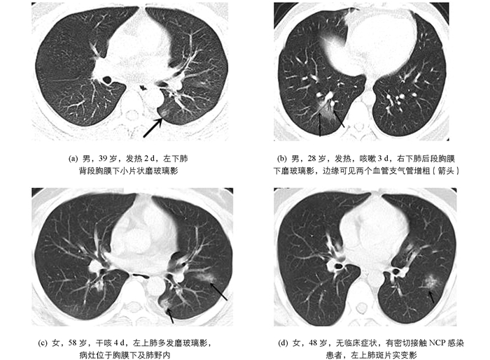
    CT阳性病灶部位分布特点(表 1):病灶位于右下肺(70/224,31.25%)及左下肺叶(68/224,30.36%)多见;病灶分布于胸膜下及肺野外带58例(58/70,82.86%),其他部位分布12例(12/70,17.14%).

  • 发病早期临床各型病灶累及肺叶数量(表 2):82例NCP患者中,属于轻型患者10例,病灶累及肺叶28个;普通型60例,病灶累及肺叶149个;重型及危重型患者12例,病灶累及肺叶37个.临床各型患者发病早期病灶累及肺叶数量比较差异无统计学意义(F=2.317,p > 0.05),两两间比较差异无统计学意义.

    临床表现:82例NCP患者中发热60例(73.17%),咳嗽52例(63.41%),多为干咳(44例,53.66%),肌肉酸痛27例(32.93%),腹痛、胸闷、恶心及纳差各10例(12.20%),无症状7例(8.54%).

3.   讨论
  • 新型冠状病毒肺炎(NCP)是严重急性呼吸综合征冠状病毒2(SARS-CoV-2)感染的疾病,主要经呼吸道飞沫和接触传播.气溶胶和消化道等传播途径尚待明确,NCP发病率高,传播迅速,是一种新发的具有较强传染性的肺部疾病[2-4]. NCP须由核酸检测或基因测序作为确诊的关键指标.然而,由于核酸检测或基因测序样本采集、运输和试剂盒性能受到诸多因素影响,导致检测不便,且核酸检测也存在假阴性可能.胸部CT作为诊断肺炎的常规影像学手段,具有操作简便、诊断速度快等优点. Ai等[5]发现,根据核酸阳性结果,胸部CT提示NCP的敏感性为97%(95%CI,95%~98%,580/601),胸部CT对NCP的诊断具有较高的敏感性,可作为目前流行区NCP检测的主要工具. 2020年3月4日,国家卫生健康委也发布了《新型冠状病毒肺炎诊疗方案(试行第七版)》,特别强调了CT影像学对于湖北防控策略“早诊断、早隔离”的重要价值.本文着重分析NCP患者早期胸部CT影像表现及临床特点,尝试尽早诊断该病,这对于疾病治疗及控制疫情具有重要意义.

    磨玻璃影(GGO)是在CT肺窗上表现为肺透亮度稍下降、肺密度稍增高的一种阴影,支气管及血管边缘仍能显示,纵隔窗GGO不显示. GGO形成原因为[6-7]:①肺泡的容积减少及含气量减少,肺泡腔内气体被渗出液、血液、炎性细胞或无定型物质置换;②肺组织间质及肺泡壁增厚,因水肿、肿瘤增生、纤维化和非癌性肉芽肿沉积而增厚;③肺栓塞及肺血管炎所致肺血流灌注相对增加.肺部GGO可以为良性病变,如局灶性间质纤维化、炎症、感染和出血,也可以是肺腺癌及浸润前病变[8].

    GGO表现多种多样,间质性肺炎表现为弥漫性GGO,多伴有蜂窝状透亮影及网格影.双肺坠积性效应有按重力分布特点,多表现在双下肺近背侧弧分布形.心源性肺水肿患者多有心、肾功能障碍,且多为双侧、对称分布,伴有小叶间均匀增厚.

    我们发现,NCP病灶呈胸膜下GGO[58(58/70,82.86%)]分布多见,可能是因为SARS-CoV-2直接侵犯终末细支气管远端的肺腺泡,导致肺泡损伤[9]并侵犯肺泡周围血管,引起胸膜下血管炎,导致肺泡内炎性渗出物增加,空气含量减少,周围肺间质及肺泡壁增厚,形成了大量胸膜下GGO及血管束增粗. Heitzman等[10]认为,肺分为周围的肺皮质和中央的肺髓质.周围的肺皮质内的肺小叶形状为扇形或弧形排列,肺外周有丰富的毛细血管及有换气功能的肺泡,易引发炎症反应,NCP患者双肺胸膜下病灶正好符合这种肺小叶解剖上的差异.而NCP患者早期病灶渗出不明显,多不伴胸腔积液及大片实变影.

    在本组病例中,我们发现双肺GGO以右下肺(70/224,31.25%)及左下肺叶(68/224,30.36%)为主,有文献认为双下肺为主,右肺中叶受累相对少见[11],这可能与肺内气流分布有关,具体原因还有待研究.

    临床各型患者发病早期病灶累及肺叶数量相比较差异无统计学意义(F=2.317,p > 0.05).武汉大学中南医院WANG等[12]报告的138例NCP住院患者中,36例重症患者有明显的基础疾病,包括高血压病(58.3%)、糖尿病(22.2%)、心血管疾病(25%)、脑血管疾病(16.7%).陈雷等[13]发现患者血清中白介素-2受体(IL-2R)、白介素-6(IL-6)水平与病情严重程度有明显的相关性.这些研究可能说明,新冠肺炎的严重程度可能与患者基础疾病有关,也可能与个体对疾病的反应有关,而与早期病灶数量累及肺叶的多少没有关系.

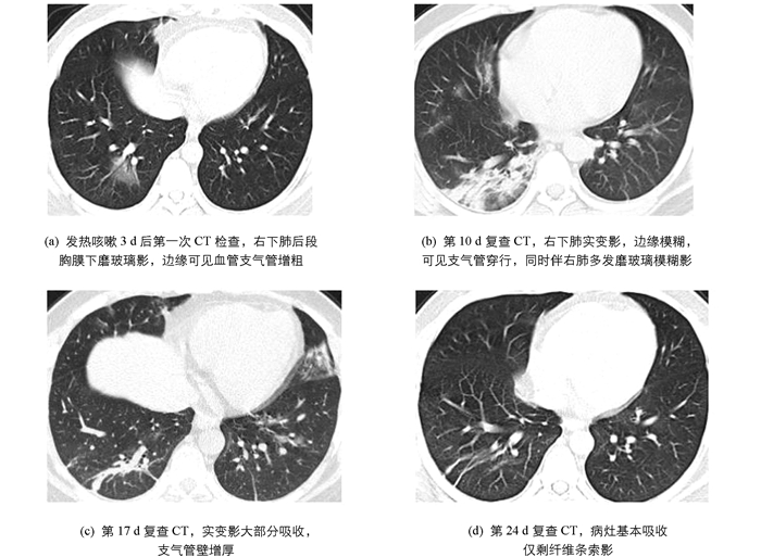
    病情发展预测:据中华医学会放射学分会专家推荐意见[14],将NCP的CT表现分为早期、进展期和重症期.早期[图 2(a)]即为单发或多发GGO;进展期[图 2(b)]病变范围进一步扩大、分布区域增多,可见实变影及空气支气管征伴小叶间隔增厚;重症期为双肺弥漫性实变,部分呈“白肺”表现.绝大多数患者经过治疗,病灶范围缩小,密度减低,实变逐渐消失,部分残留条索灶[图 2(c)(d)].

    目前也是流感的高发季节,在当前特殊时期若发现肺内有GGO、实变、结节等影像表现,需要排除病毒性肺炎、重症急性呼吸窘迫综合征(SRARS)、支原体及细菌感染等.甲型流感病毒肺炎多是单侧或双侧局灶或多发GGO,伴或不伴实变,常沿支气管血管束分布或胸膜下分布[15-16],NCP早期可表现为小GGO或小片磨玻璃影内见增粗血管影,可能有助鉴别.腺病毒肺炎常见于儿童,胸部CT表现为双肺弥漫或局限性GGO,伴或不伴实变. SRARS与NCP致病病毒同属冠状病毒,两者致病机制和影像学表现高度相似,单纯影像学难以鉴别,最终需借助病原学检测.支原体肺炎儿童和青少年常见,最常见的症状有发热、刺激性干嗽.胸部CT呈沿支气管血管束分布为主的气腔实变及GGO,呈自肺门向肺野外围伸展的大片扇形阴影,可见支气管壁增厚及细支气管“树芽征”与肺门及纵隔淋巴结肿大,实验室检查支原体抗体阳性[17].细菌性肺炎多无上呼吸道感染前驱症状,咳脓性痰,血性痰或铁锈色痰,白细胞数增高,影像学多表现为肺叶或段实变影,抗生素治疗效果好.

    总之,NCP有其特有的影像学征象,CT阳性患者早期主要表现为胸膜下和/或双肺野外带磨玻璃影及斑片影,常合并灶内血管束增粗,临床有发热、咳嗽、肌肉酸痛等症状,实验室检查白细胞计数减少、淋巴细胞比例降低,C反应蛋白上升,且有流行病学史时需考虑NCP.确诊NCP的金标准为核酸检测阳性,对于核酸检测阴性及CT检查呈阴性的患者,也不能大意,临床应及时复查,以便早发现、早诊断、早隔离、早治疗.

Figure (2)  Table (2) Reference (17)

Catalog

    /

    DownLoad:  Full-Size Img  PowerPoint
    Return
    Return