Message Board

Dear readers, authors and reviewers,you can add a message on this page. We will reply to you as soon as possible!

2017 Volume 39 Issue 9
Article Contents

Yan-jiao PANG, Yuan-yuan YU, Hu-zhi ZHENG, et al. Colorimetric Detection of Glutathione Based on Peroxidase-Like Activity of Papain[J]. Journal of Southwest University Natural Science Edition, 2017, 39(9): 125-130. doi: 10.13718/j.cnki.xdzk.2017.09.019
Citation: Yan-jiao PANG, Yuan-yuan YU, Hu-zhi ZHENG, et al. Colorimetric Detection of Glutathione Based on Peroxidase-Like Activity of Papain[J]. Journal of Southwest University Natural Science Edition, 2017, 39(9): 125-130. doi: 10.13718/j.cnki.xdzk.2017.09.019

Colorimetric Detection of Glutathione Based on Peroxidase-Like Activity of Papain

More Information
  • Corresponding author: Yi-juan LONG
  • Received Date: 22/11/2016
    Available Online: 20/09/2017
  • MSC: O657

  • Papain, a newly discovered proteinase, possesses peroxidase enzyme activity, which can catalyze the oxidation of the peroxidase substrate 3, 3', 5, 5'-tetramethylbenzidine (TMB) in the presence of H2O2 to the oxidized TMB to induce a blue colored reaction with a typical absorption peak centered at 652 nm. However, GSH can consume H2O2, which results in a fading of the blue color of the reaction system and a decrease of the absorbance. Accordingly, GSH can be detected by measuring the wastage of H2O2. Based on this approach, a facile, sensitive and selective colorimetric assay has been developed to detect GSH, with a promising application in biological fluids.
  • 加载中
  • [1] NI P, SUN Y, DAI H, et al. Highly Sensitive and Selective Colorimetric Detection of Glutathione Based on Ag [J] ion-3, 3', 5, 5'-Tetramethylbenzidine (TMB) [J]. Biosensors and Bioelectronics, 2015, 63: 47-52. doi: 10.1016/j.bios.2014.07.021

    CrossRef Google Scholar

    [2] KRAUTH-SIEGEL R L, BAUER H, SCHIRMER R H. Dithiol Proteins as Guardians of the Intracellular Redox Milieu in Parasites: Old and New Drug Targets in Trypanosomes and Malaria-Causing Plasmodia [J]. Angewandte Chemie International Edition, 2005, 44(5): 690-715. doi: 10.1002/(ISSN)1521-3773

    CrossRef Google Scholar

    [3] LI M, WU X, WANG Y, et al. A Near-Infrared Colorimetric Fluorescent Chemodosimeter for the Detection of Glutathione in Living Cells [J]. Chemical Communications, 2014, 50(14): 1751-1753. doi: 10.1039/c3cc48128j

    CrossRef Google Scholar

    [4] SCHIRMER R H, MVLLER J G, KRAUTH-SIEGEL R L. Disulfide-Reductase Inhibitors as Chemotherapeutic Agents: The Design of Drugs for Trypanosomiasis and Malaria [J]. Angewandte Chemie International Edition in English, 1995, 34(2): 141-154. doi: 10.1002/(ISSN)1521-3773

    CrossRef Google Scholar

    [5] SHI Y, PAN Y, ZHANG H, et al. A Dual-Mode Nanosensor Based on Carbon Quantum Dots and Gold Nanoparticles for Discriminative Detection of Glutathione in Human Plasma [J]. Biosensors and Bioelectronics, 2014, 56: 39-45. doi: 10.1016/j.bios.2013.12.038

    CrossRef Google Scholar

    [6] BAYRAM B, RIMBACH G, FRANK J, et al. Rapid Method for Glutathione Quantitation Using High-Performance Liquid Chromatography with Coulometric Electrochemical Detection [J]. Journal of Agricultural and Food Chemistry, 2013, 62(2): 402-408.

    Google Scholar

    [7] GAO X, LI X, LI L, et al. A Simple Fluorescent Off-On Probe for the Discrimination of Cysteine from Glutathione [J]. Chemical Communications, 2015, 51(45): 9388-9390. doi: 10.1039/C5CC02788H

    CrossRef Google Scholar

    [8] TSARDAKA E C, ZACHARIS C K, TZANAVARAS P D, et al. Determination of Glutathione in Baker's Yeast by Capillary Electrophoresis Using Methyl Propiolate as Derivatizing Reagent [J]. Journal of Chromatography A, 2013, 1300: 204-208. doi: 10.1016/j.chroma.2013.05.005

    CrossRef Google Scholar

    [9] MIAO P, LIU L, NIE Y, et al. An Electrochemical Sensing Strategy for Ultrasensitive Detection of Glutathione by Using Two Gold Electrodes and Two Complementary Oligonucleotides [J]. Biosensors and Bioelectronics, 2009, 24(11): 3347-3351. doi: 10.1016/j.bios.2009.04.041

    CrossRef Google Scholar

    [10] 徐秀芳. 纳米材料模拟酶及其分析应用[D]. 无锡: 江南大学, 2014.http://cdmd.cnki.com.cn/Article/CDMD-10295-1014380840.htm

    Google Scholar

    [11] KAUL P, SATHISH H A, PRAKASH V. Effect of Metal Ions on the Structure and Activity of Papain from Carcia Papaya [J]. Nahrung/Food, 2002, 46(1): 2-6. doi: 10.1002/1521-3803(20020101)46:1<2::AID-FOOD2>3.0.CO;2-1

    CrossRef Google Scholar

    [12] 于缓缓. 木瓜蛋白酶过氧化物模拟酶性质及其分析应用[D]. 重庆: 西南大学, 2016.http://cdmd.cnki.com.cn/Article/CDMD-10635-1016766856.htm

    Google Scholar

    [13] HAN B, YUAN J, WANG E. Sensitive and Selective Sensor for Biothiols in the Cell Based on the Recovered Fluorescence of the CdTe Quantum Dots? Hg (II) system [J]. Analytical Chemistry, 2009, 81(13): 5569-5573. doi: 10.1021/ac900769h

    CrossRef Google Scholar

    [14] LIU J, BAO C, ZHONG X, et al. Highly Selective Detection of Glutathione Using Aquantum-Dot-Based OFF-ON Fluorescent Probe [J]. Chemical Communications, 2010, 46(17): 2971-2973. doi: 10.1039/b924299f

    CrossRef Google Scholar

    [15] ZHANG N, QU F, LUO H Q, et al. Sensitive and Selective Detection of Biothiols Based on Target-Induced Agglomeration of Silver Nanoclusters [J]. Biosensors and Bioelectronics, 2013, 42: 214-218. doi: 10.1016/j.bios.2012.10.090

    CrossRef Google Scholar

    [16] HUANG Z, PU F, LIN Y, et al. Modulating DNA-Templated Silver Nanoclusters for Fluorescence Turn-On Detection of Thiol Compounds [J]. Chemical. Communications, 2011, 47(12), 3487-3489. doi: 10.1039/c0cc05651k

    CrossRef Google Scholar

    [17] SHAMSIPUR M, SAFAVI A, MOHAMMADPOUR Z. Indirect Colorimetric Detection of Glutathione Based on Its Radical Restoration Ability Using Carbon Nanodots as Nanozymes [J]. Sensors and Actuators B: Chemical, 2014, 199: 463-469. doi: 10.1016/j.snb.2014.04.006

    CrossRef Google Scholar

    [18] CHEN X Q, KO S K, KIM M J, et al. A Thiol-Specific Fluorescent Probe and Its Application for Bioimaging [J]. Chemical Communications, 2010, 46(16): 2751-2753. doi: 10.1039/b925453f

    CrossRef Google Scholar

    [19] MCDERMOTT G P, FRANCIS P S, HOLT K J, et al. Determination of Intracellular Glutathione and Glutathione Disulfide Using High Performance Liquid Chromatography with Acidic Potassium Permanganate Chemiluminescence Detection [J]. Analyst, 2011, 136(12): 2578-2585. doi: 10.1039/c1an00004g

    CrossRef Google Scholar

  • 加载中
通讯作者: 陈斌, bchen63@163.com
  • 1. 

    沈阳化工大学材料科学与工程学院 沈阳 110142

  1. 本站搜索
  2. 百度学术搜索
  3. 万方数据库搜索
  4. CNKI搜索

Figures(4)  /  Tables(2)

Article Metrics

Article views(1317) PDF downloads(938) Cited by(0)

Access History

Other Articles By Authors

Colorimetric Detection of Glutathione Based on Peroxidase-Like Activity of Papain

    Corresponding author: Yi-juan LONG

Abstract: Papain, a newly discovered proteinase, possesses peroxidase enzyme activity, which can catalyze the oxidation of the peroxidase substrate 3, 3', 5, 5'-tetramethylbenzidine (TMB) in the presence of H2O2 to the oxidized TMB to induce a blue colored reaction with a typical absorption peak centered at 652 nm. However, GSH can consume H2O2, which results in a fading of the blue color of the reaction system and a decrease of the absorbance. Accordingly, GSH can be detected by measuring the wastage of H2O2. Based on this approach, a facile, sensitive and selective colorimetric assay has been developed to detect GSH, with a promising application in biological fluids.

  • 谷胱甘肽(Glutathione,GSH),即γ-L-谷氨酰-L-半胱氨酰-甘氨酸,是一种由L-谷氨酸、L-半胱氨酸和甘氨酸经肽键缩合而成的生物活性三肽,也是一种广泛存在于生物体内的非蛋白硫醇化合物[1].自然界中谷胱甘肽以还原型(GSH)和氧化型(GSSG)2种状态存在,通常所说的谷胱甘肽是指还原型谷胱甘肽. GSH的主要特点是具有游离的巯基和很强的供电子或质子的能力,它可以作为水相的抗氧化剂和抗氧化酶的辅因子.这些结构特点决定了在生物体内发挥重要的生理功能:细胞内氧化还原活性的维护[2]、细胞内信号转换[3]、细胞的代谢[4]、基因调控[5].同时,GSH也广泛应用于医药、食品和化妆品行业.

    目前,关于GSH的检测方法较多,如高效液相色谱法[6]、荧光法[7]、毛细管电泳法[8]和电化学检测法[9]等.对于GSH的检测,虽然报道的光学生物传感方法大部分比较简单,有较高灵敏度,但存在实验步骤繁琐、样品处理耗时等缺点.近年来,随着纳米技术的不断发展,纳米微粒作为模拟酶分析应用的研究引起了广泛关注.与天然酶相比,纳米酶具有一些优点:低成本的可控合成、高的催化活性、能够耐受更苛刻的环境等[10].但是,这些纳米材料的制备程序和修饰步骤复杂耗时,且在合成过程中容易团聚,合成的纳米材料也存在批间差异,从而使结果重现性低,本研究发现一种生物催化剂——木瓜蛋白酶具有类似过氧化物酶的性质.基于此,开发了一种新型的GSH检测方法来满足临床、生物和医疗需求.

    木瓜蛋白酶(EC3.4.22.2) 是木瓜中含有的一种低特异性蛋白水解酶,属巯基蛋白酶,由212个氨基酸残基组成的一条肽链,活性位点为25位的半胱氨酸残基和158位的组氨酸残基[11].它具有耐高温、稳定性好和蛋白水解能力强等特征,在食品和医药等领域有广泛的应用.最近笔者所在研究组发现木瓜蛋白酶具有类似过氧化物酶的性质,能催化H2O2氧化过氧化物酶底物并产生特定的颜色反应[12].而本研究发现,GSH可以抑制木瓜蛋白酶催化TMB-H2O2显色反应.基于此现象,本研究建立了快速简单的方法用来高灵敏和高选择地检测GSH,该方法操作简单,不需要复杂的仪器设备,并成功用于实际样品中GSH的检测.

1.   实验部分
  • H2O2(30%)、磷酸氢二钠、磷酸二氢钠、葡萄糖、抗坏血酸、氢氧化钠、氯化钠、氯化钾、硫酸镁、硝酸钙、硫酸锌、硫酸铝、硫酸亚铁和硫酸铜购自重庆川东化工有限公司;3,3',5,5'-四甲基联苯胺(TMB)、丙氨酸(Ala)、异亮氨酸(Ile)、酪氨酸(Tyr)、甲硫氨酸(Met)、天门冬氨酸(Asp)、精氨酸(Arg)、缬氨酸(Val)、赖氨酸(Lys)、组氨酸(His)、亮氨酸(Leu)、苯丙氨酸(Phe)、谷氨酸(Glu)、甘氨酸(Gly)、胱氨酸(Cystime)和GSH购自上海生工生物工程有限公司;超滤管(截留分子量为5 kDa)购自Sigma-Aldrich公司;尿样为西南大学校医院提供;所有试剂均为分析纯,可直接使用;本实验用水均为Milli-Q-Plus超纯水系统所制备的超纯水(Millipore,18.2 MΩ,美国).

    UV-2450紫外可见分光光度计(日本岛津公司);漩涡混合器(金坛市医疗仪器厂);高速台式冷冻离心机LRH-250-Z383K(德国HERMLE公司);电热恒温水浴槽(上海比朗仪器有限公司).

  • 首先,配置10 mmol/L的GSH储备液,并通过梯度稀释获得不同浓度的GSH.再将200 μL 1.0 μg/mL木瓜蛋白酶与不同浓度的GSH于35 ℃孵育30 min,然后向混合液中分别加入6.0 mmol/L TMB,10 mmol/L H2O2和0.1 mol/L磷酸盐缓冲溶液(pH=5.0)200 μL,并加水至终体积为2 mL;混合均匀后,35 ℃继续孵育2 h,用紫外可见分光光度计测其在652 nm的吸光度.对于实际样品的检测,先将样品以超滤管3 000 r/min离心30 min,滤液以超纯水稀释一定倍数,代替上述的谷胱甘肽标准液,按同样的方法进行测定.

2.   结果与讨论
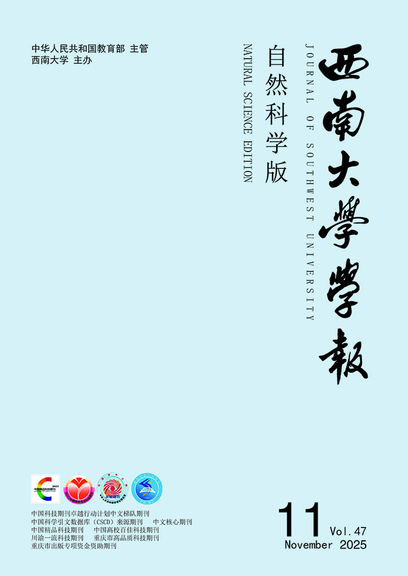
  • 研究发现木瓜蛋白酶具有过氧化物酶活性,能催化H2O2氧化过氧化物酶底物TMB,产生蓝色反应,并在652 nm处有吸收峰(图 1);而GSH会消耗H2O2,导致吸光度值降低且溶液颜色变浅.基于此,本研究设计了一种比色分析法检测GSH,其检测原理为:

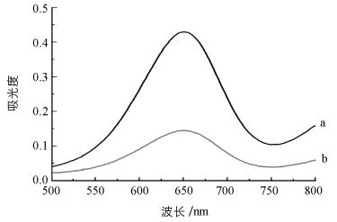
  • 实验中优化了pH值、温度、H2O2浓度及TMB浓度对检测GSH的影响(图 2). 图 2中纵坐标ΔA=A0-A,其中A0A分别表示在不加GSH和加入GSH的吸光度值.在1.0~8.0的范围内优化了pH值(图 2(a)):pH值从1.0逐渐增大时,ΔA也随之增大;当pH进一步增大时,ΔA逐渐降低.因此,5.0为最佳pH值.在20~50 ℃范围内优化了最佳温度:孵育温度在25~35 ℃之间,ΔA几乎没有差别;然而当温度超过35 ℃时,随着温度的升高,ΔA值降低(图 2(b)).因此,选择35 ℃为最佳反应温度.当H2O2浓度在0.10~1.0 mmol/L范围内时,ΔA值随着H2O2浓度的增大而增大;在1.0~8.0 mmol/L范围内,ΔA随着浓度的增大而降低,这是因为较高浓度的H2O2会抑制木瓜蛋白酶的催化活性(图 3(c)).因此,选择1.0 mmol/LH2O2浓度为最佳测定条件.在0.10~1.0 mmol/L范围内优化了TMB浓度(图 2(d)),当TMB浓度为0.60 mmol/L时,ΔA值达到最大,因此选择TMB浓度为0.60 mmol/L.

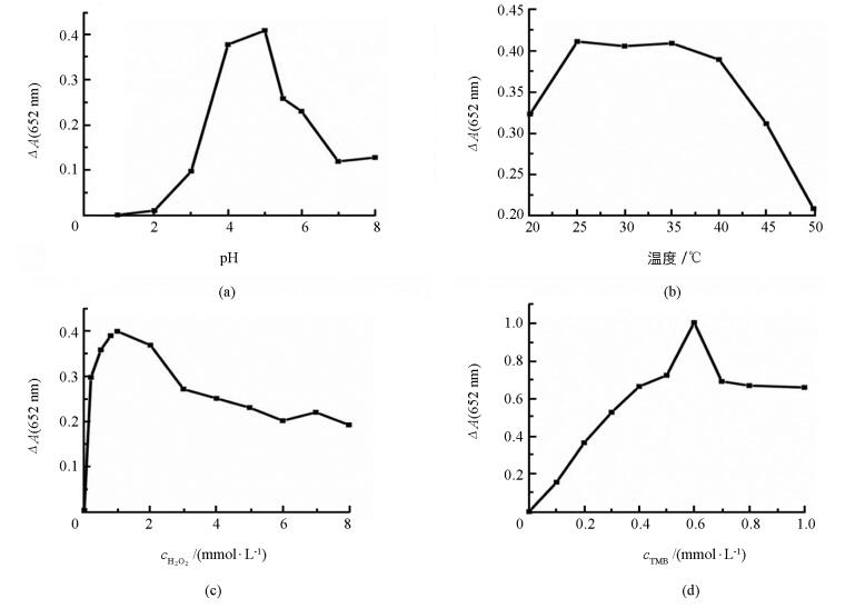
  • 在最佳的实验条件下,将不同浓度的GSH加入反应体系中,通过检测木瓜蛋白酶-TMB-H2O2反应体系在652 nm的吸光度,实现对GSH的检测.对应光谱图和标准曲线(图 3),在0.10~10 μmol/L范围内,GSH浓度与吸光度具有良好的线性关系,其线性方程为ΔA=0.017 6c+0.190 9,相关系数为0.991 2,检出限为0.03 μmol/L.与其他方法比较(表 1),本方法具有较高的灵敏度.此外,本实验不需要合成纳米材料,也不需要复杂的仪器设备,且操作过程简单,表明该方法在生物技术、生物分析及生物医学上有潜在应用.

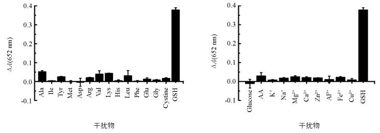
  • 为了考察该方法的选择性,向木瓜蛋白酶-TMB-H2O2显色体系中分别加入5 μmol/L的葡萄糖、抗坏血酸、14种氨基酸(丙氨酸、异亮氨酸、酪氨酸、甲硫氨酸、天门冬氨酸、精氨酸、缬氨酸、赖氨酸、组氨酸、亮氨酸、苯丙氨酸、谷氨酸、甘氨酸、胱氨酸)、8种金属离子(K+,Na+,Mg2+,Ca2+,Zn2+,Al3+,Fe2+,Cu2+)等常见的干扰物,并在上述最佳条件下,测其在652 nm的吸光度(图 4).结果显示,同浓度的干扰物质均对显色体系无明显影响,表明本方法对GSH有很高的选择性.

  • 为了证实该方法在实际应用中的可行性,检测了人尿液样品中GSH的含量.将尿液样品超滤处理后,并稀释至一定倍数,采用标准加入法进行检测,所得回收率为97%~109.4%,结果列于表 2中.表明该方法能够成功用于实际样品中GSH的分析检测.

3.   结论
  • 基于木瓜蛋白酶的过氧化物酶活性设计了一种简便、灵敏地检测GSH浓度的比色分析法.木瓜蛋白酶可以催化H2O2氧化TMB产生蓝色反应,并在652 nm处有吸收峰;而当加入GSH后,由于GSH会消耗H2O2,导致在652 nm处的吸收峰强度降低. GSH检测的线性范围为0.10~10 μmol/L,检出限为0.03 μmol/L.该方法具有灵敏、简单、成本低廉且选择性良好的优点,可用于实际样品的检测.

Figure (4)  Table (2) Reference (19)

Catalog

    /

    DownLoad:  Full-Size Img  PowerPoint
    Return
    Return