-
犬弓首蛔虫Toxocara canis,T. canis是犬常见的胃肠道寄生线虫,其感染性幼虫可以感染人和多种动物[1],是引起弓首蛔虫病的主要病原.该病的流行和分布极为广泛,据资料报道其感染率为1.4%~64.7%[2-3]. T. canis感染人和其他非特异性宿主可引起内脏幼虫移行症(Visceral larva migrans,VLM)、眼睛幼虫移行症(Ocular larva migrans,OLM)和神经幼虫移行症(Neurological larva migrans,NLM),造成严重的病理综合征[4-7].
卵黄原蛋白(vitellogenin,Vg)是脂质转运蛋白家族的成员之一[8-9],普遍存在于卵生非哺乳动物体内[10].不同来源的卵黄原蛋白具有3个保守功能结构域:位于N端的功能结构域(Vitellogenin,LPD_N)、未知结构域(domain of unknow function,DUF1943)和位于C端的vWD结构域(von-Willebrand factor type D domain,vWD)[11-13]. Vg主要是在卵巢外组织中合成,分泌进入循环系统到达卵巢,通过受体介导的内吞作用进入卵母细胞,为正在发育的胚胎或幼虫提供营养物质[14-15].此外,Vg还具有免疫防御、粒子载体、抗氧化活性、筛选内分泌干扰物、调查环境毒理和污染等[16-19]作用.目前,对于昆虫、甲壳动物和脊椎动物的Vg研究较多[20-21],但对于寄生虫卵黄原蛋白基因的研究相对较少.
本研究拟运用分子生物学技术,克隆犬弓首蛔虫Tc-duf1943,构建pET-28a(+)/duf1943原核表达载体,以期能在大肠杆菌E. coli中得到表达,优化其表达条件,为进一步研究TcDUF1943的生物学功能奠定基础.
Cloning and Prokaryotic Expression of the DUF1943 Domain of Toxocara canis Vitellogenin
-
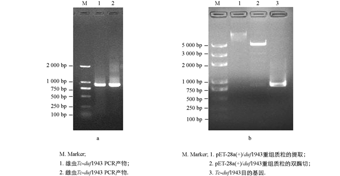
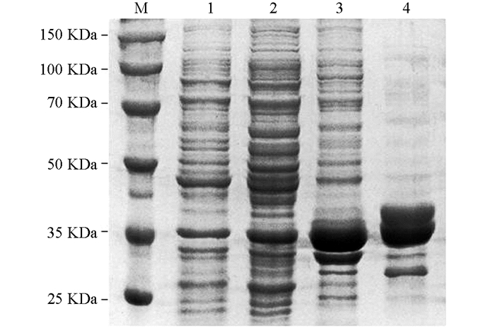
摘要: 为克隆犬弓首蛔虫Toxocara canis,T. canis卵黄原蛋白DUF1943结构域(The domain of unknown function 1943,DUF1943)(Tc-duf1943),构建其原核表达载体pET-28a(+)/duf1943,并检测重组蛋白在大肠杆菌Escherichia coli,E. coli中的表达形式.该文根据T. canis的转录组数据,以T. canis雌、雄虫cDNA为模板扩增Tc-duf1943,然后与表达载体pET-28a(+)连接,转化E. coli BL21(DE3),经IPTG诱导,对目的蛋白的原核表达形式和蛋白质纯化进行鉴定.结果显示Tc-duf1943序列长度为864 bp,含有一个完整的开放阅读框,编码288个氨基酸;SDS-PAGE电泳结果表明,重组蛋白以包涵体形式存在,相对分子质量大小约为3.5×10⁴;对表达条件进行优化后,以0.4 mmol/L IPTG,于37 ℃诱导4 h时可获得大量目的蛋白;利用Ni-NTA亲和层析柱纯化可以获得具有较高纯度的目的蛋白.该试验成功构建了pET-28a(+)/duf1943原核表达载体,为进一步研究TcDUF1943的生物学功能奠定了基础.Abstract: To clone the DUF1943 (the domain of unknown function 1943) of vitellogenin in Toxocara canis, the prokaryotic expression vector pET-28a (+) /duf1943 of Tc-duf1943 was constructed and its expression form in Escherichia coli was detected. According to the transcriptome data about T. canis, and using the cDNA of female and male as templates, Tc-duf1943 was cloned successfully. Then the target gene was connected with the prokaryotic expression vector pET-28a(+) (pET-28a(+)/duf1943), E. coli BL21 (DE3) was transformed and the prokaryotic expression and purification of the protein was identified. The results showed that Tc-duf1943 was 864 bp in length, with a complete open reading frame, encoding 288 amino acids, and SDS-PAGE indicated that the recombinant protein was expressed in inclusion bodies, with a molecular weight of about 3.5×10⁴, The optimal condition of prokaryotic expression was induced by 0.4 mmol/L IPTG for 4 hours at 37 ℃. Purified with Ni-NTA affinity chromatography, target protein with high purity was obtained. In conclusion, this experiment successfully constructed the prokaryotic expression vector of pET-28a(+)/duf1943, which laid a foundation for further biological function study of TcDUF1943.
-
Key words:
- Toxocara canis /
- DUF1943 /
- clone /
- prokaryotic expression .
-
表 1 引物序列
基因名称 上游引物(5'-3') 下游引物(5'-3') Tc-duf1943 CGCGGATCCATGAGCAAATTCATGCATAGGAC CCCAAGCTTTTAAGTTTGTATCAACAGGATCT 18S rRNA AATTGTTGGTCTTCAACGAGGA AAAGGGCAGGGACGTAGTCAA -
[1] SMITH H, HOLLAND C, TAYLOR M, et al. How Common is Human Toxocariasis? Towards Standardizing Our Knowledge [J]. Trends Parasitol, 2009, 25(4): 182-188. doi: 10.1016/j.pt.2009.01.006 [2] doi: http://www.magonlinelibrary.com/doi/pdfplus/10.12968/coan.2014.19.9.465 SCHNIEDER T, LAABS E M, WELZ C. Larval Development of Toxocara canis in Dogs [J]. Vet Parasitol, 2011, 175(3/4): 193-206. [3] doi: http://www.ncbi.nlm.nih.gov/pubmed/19091472 DAI R S, LI Z Y, LI F, et al. Severe Infection of Adult Dogs with Helminths in Hunan Province, China Poses Significant Public Health Concerns [J]. Vet Parasitol, 2009, 160(3/4): 348-350. [4] AKUTHOTA P, WELLER P F. Eosinophilic Pneumonias [J]. Clin Microbiol Rev, 2012, 25(4): 649-660. doi: 10.1128/CMR.00025-12 [5] doi: http://www.ncbi.nlm.nih.gov/pubmed/14628955 VIDAL J E, SZTAJNBOK J, SEGURO A C. Eosinophilic Meningoencephalitis Due to Toxocara canis: Case Report and Review of the Literature [J]. Am J Trop Med Hyg, 2003, 69(3): 341-343. [6] WIS ′ NIEWSKA-LIGIER M, WOZ ′ NIAKOWSKA-GESICKA T, SOBOLEWSKA-DRYJAN ′ SKA J, et al. Analysis of the Course and Treatment of Toxocariasis in Children-a Long-Term Observation [J]. Parasitol Res, 2012, 110(6): 2363-2371. doi: 10.1007/s00436-011-2772-y [7] CHEN J, XU M J, ZHOU D H, et al. Canine and Feline Parasitic Zoonoses in China [J]. Parasit Vectors, 2012, 5(1): 152-160. doi: 10.1186/1756-3305-5-152 [8] SMOLENAARS M M, MADSEN O, RODENBURG K W, et al. Molecular Diversity and Evolution of the Large Lipid Transfer Protein Superfamily [J]. J Lipid Res, 2007, 48(3): 489-502. doi: 10.1194/jlr.R600028-JLR200 [9] BABIN P J, GIBBONS G F. The Evolution of Plasma Cholesterol: Direct Utility or a "Spandrel" of Hepatic Lipid Metabolism? [J]. Prog Lipid Res, 2009, 48(2): 73-91. doi: 10.1016/j.plipres.2008.11.002 [10] doi: https://www.sciencedirect.com/science/article/pii/S0378111900003760 WANG H, YAN T, TAN J T, et al. A Zebrafish Vitellogenin Gene (vg3) Encodes a Novel Vitellogenin Without a Phosvitin Domain and May Represent a Primitive Vertebrate Vitellogenin Gene [J]. Gene, 2000, 256(1/2): 303-310. [11] AVARRE J C, LUBZENS E, BABIN P J. Apolipocrustacein, Formerly Vitellogenin, is the Major Egg Yolk Precursor Protein in Decapod Crustaceans and is Homologous to Insect Apolipophorin Ⅱ/Ⅰ and Vertebrate Apolipoprotein B [J]. BMC Evol Biol, 2007, 7(1): 3-13. doi: 10.1186/1471-2148-7-3 [12] TUFAIL M, TAKEDA M. Insect Vitellogenin/Lipophorin Receptors: Molecular Structures, Role in Oogenesis, and Regulatory Mechanisms [J]. J Insect Physiol, 2009, 55(2): 87-103. doi: 10.1016/j.jinsphys.2009.01.009 [13] SAWAGUCHI S, KOYA Y, YOSHIZAKI N, et al. Multiple Vitellogenins (Vgs) in Mosquitofish (Gambusia affinis): Identification and Characterization of Three Functional Vg Genes and Their Circulating and Yolk Protein Products [J]. Biol Reprod, 2005, 72(4): 1045-1060. doi: 10.1095/biolreprod.104.037895 [14] ARUKWE A, GOKSØYR A. Eggshell and Egg Yolk Proteins in Fish: Hepatic Proteins for the Next Generation: Oogenetic, Population, and Evolutionary Implications of Endocrine Disruption [J]. Comp Hepatol, 2003, 2(1): 4-24. doi: 10.1186/1476-5926-2-4 [15] REITH M, MUNHOLLAND J, KELLY J, et al. Lipovitellins Derived from Two Forms of Vitellogenin Are Differentially Processed During Oocyte Maturation in Haddock (Melanogrammus aeglefinus) [J]. J Exp Zool, 2001, 291(1): 58-67. doi: 10.1002/(ISSN)1097-010X [16] SEEHUUS S C, NORBERG K, GIMSA U, et al. Reproductive Protein Protects Functionally Sterile Honey Bee Workers from Oxidative Stress [J]. Proc Natl Acad Sci USA, 2006, 103(4): 962-967. doi: 10.1073/pnas.0502681103 [17] GERBRON M, GERAUDIE P, XUEREB B, et al. In vitro and in Vivo Studies of the Endocrine Disrupting Potency of Cadmium in Roach (Rutilus rutilus) liver [J]. Mar Pollut Bull, 2015, 95(2): 582-589. doi: 10.1016/j.marpolbul.2015.03.043 [18] SERRANO M L, LUQUE M E, SÁNCHEZ S S. Xepac Protein and IP3/Ca2+ Pathway Implication During Xenopus laevis Vitellogenesis [J]. Zygote, 2015, 23(1): 99-110. doi: 10.1017/S0967199413000324 [19] ZHANG S, WANG S, LI H, et al. Vitellogenin, a Multivalent Sensor and an Antimicrobial Effector [J]. Int J Biochem Cell Biol, 2011, 43(3): 303-305. doi: 10.1016/j.biocel.2010.11.003 [20] JEON J M, LEE S O, KIM K S, et al. Characterization of Two Vitellogenin cDNAs from a Pandalus Shrimp (Pandalopsis japonica): Expression in Hepatopancreas is Down-Regulated by Endosulfan Exposure [J]. Comp Biochem Physiol B Biochem Mol Biol, 2010, 157(1): 102-112. doi: 10.1016/j.cbpb.2010.05.006 [21] JIA X, CHEN Y, ZOU Z, et al. Characterization and Expression Profile of Vitellogenin Gene from Scylla paramamosain [J]. Gene, 2013, 520(2): 119-130. doi: 10.1016/j.gene.2013.02.035 [22] doi: http://onlinelibrary.wiley.com/doi/10.1111/j.1365-2109.2009.02220.x/full FINN R N, FYHN H J. Requirement for Amino Acids in Ontogeny of Fish [J]. Aquacult Res, 2010, 41(5): 684-716. [23] PIULACHS M D, GUIDUGLI K R, BARCHUK A R, et al. The Vitellogenin of the Honey Bee, Apis mellifera: Structural Analysis of the cDNA and Expression Studies [J]. Insect Biochem Mol Biol, 2003, 33(4): 459-465. doi: 10.1016/S0965-1748(03)00021-3 [24] MATOZZO V, GAGNÉ F, MARIN M G, et al. Vitellogenin as a Biomarker of Exposure to Estrogenic Compounds in Aquatic Invertebrates: a Review [J]. Environ Int, 2008, 34(4): 531-545. doi: 10.1016/j.envint.2007.09.008 [25] KANG B J, JUNG J H, LEE J M, et al. Structural and Expression Analyses of Two Vitellogenin Genes in the Carp, Cyprinus carpio [J]. Comp Biochem Physiol B Biochem Mol Biol, 2007, 148(4): 445-453. doi: 10.1016/j.cbpb.2007.07.088 [26] 马鑫, 王斌, 胡雨晴, 等.梁平柚葡萄糖基转移酶基因的克隆及原核表达[J].西南大学学报(自然科学版), 2011, 33(8): 63-67. doi: http://xbgjxt.swu.edu.cn/jsuns/jsuns/ch/reader/view_abstract.aspx?file_no=xnnydxxb201108014&flag=1 [27] doi: https://www.sciencedirect.com/science/article/pii/007668799085008C STUDIER W F, ROSENBERG A H, DUNN J J, et al. Use of T7 RNA Polymerase to Direct Expression of Cloned Genes [J]. Methods Enzymol, 1990, 185(1): 60-89. [28] LI Z J, ZHANG S C, LIU Q H. Vitellogenin Functions as a Multivalent Pattern Recognition Receptor with an Opsonic Activity [J]. PLoS ONE, 2008, 3(4): e1940. doi: 10.1371/journal.pone.0001940 [29] LI Z, ZHANG S, ZHANG J, et al. Vitellogenin is a Cidal Factor Capable of Killing Bacteria via Interaction with Lipopolysaccharide and Lipoteichoic Acid [J]. Mol Immunol, 2009, 46(16): 3232-3239. doi: 10.1016/j.molimm.2009.08.006 [30] KHALIL S M S, DONOHUE K V, THOMPSON D M, et al. Full-Length Sequence, Regulation and Developmental Studies of a Second Vitellogenin Gene from the American Dog Tick, Dermacentor Variabilis [J]. Insect Physiol, 2011, 57(3): 400-408. doi: 10.1016/j.jinsphys.2010.12.008 [31] DU X Y, WANG X, WANG S, et al. Functional Characterization of Vitellogenin_N Domain, Domain of Unknown Function 1943, and von Willebrand Factor Type D Domain in Vitellogenin of the Non-Bilaterian Coral Euphyllia ancora: Implications for Emergence of Immune Activity of Vitellogenin in Basal Metazoan [J]. Dev Comp Immunol, 2017, 67(2): 485-494.