Message Board

Dear readers, authors and reviewers,you can add a message on this page. We will reply to you as soon as possible!

2023 Volume 45 Issue 5
Article Contents

LI Weifen, HE Pengbo, CHEN Hongmei, et al. Morphological and Molecular Identification of the Pathogen Causing Wax Apple Anthracnose in Southern Yunnan, China[J]. Journal of Southwest University Natural Science Edition, 2023, 45(5): 83-91. doi: 10.13718/j.cnki.xdzk.2023.05.008
Citation: LI Weifen, HE Pengbo, CHEN Hongmei, et al. Morphological and Molecular Identification of the Pathogen Causing Wax Apple Anthracnose in Southern Yunnan, China[J]. Journal of Southwest University Natural Science Edition, 2023, 45(5): 83-91. doi: 10.13718/j.cnki.xdzk.2023.05.008

Morphological and Molecular Identification of the Pathogen Causing Wax Apple Anthracnose in Southern Yunnan, China

More Information
  • Corresponding author: TAN Wanzhong
  • Received Date: 21/05/2022
    Available Online: 20/05/2023
  • MSC: Q949.762.2

  • Wax apple (Syzygium samarangense) is an important and high-economic value fruit tree planted broadly in the tropic and subtropic countries. The anthracnose is a fungal disease that is widespread in the wax-apple growing areas and usually causes serious yield reductions. In 2021, wax apple anthracnose was observed during the surveys of diseases of tropical fruit trees in Sipsongpanna and Pu'er, Yunnan, China and found that the disease was widespread and serious. Therefore, the present study was conducted to investigate the symptom of disease, isolate and identify the pathogen of disease. Typical spot of the anthracnose on wax apple leaves observed in this study was oval or irregular, with a gray-white center surrounded by a brown band and yellow halo. Later, the central necrotic tissue was dried and fallen, leaving holes on the leaf. The pathogen of the disease also infected the fruits and caused fruit rot during storage. Seven fungal isolates were obtained from diseased leaf tissues. Laboratory tests with Koch's Postulates verified that LWTJ2 from Pu'er and LB4 from Sipsongpanna was the pathogen causing wax apple anthracnose. The pathogenic isolates were morphologically indistinguishable. After cultured on potato glucose agar (PDA) medium plates at 25℃ for 6 d, the colonies were round with complete edges and prosperous aerial hyphae, surface smooth and villous, white or light-yellow colored. At 12 d after cultivation, the colony became pale-white with gray-black center, producing plenty of orange-colored conidia. Under microscope, the hyphae were hyaline, septate, and branched at near right-angles. Conidia were colorless, long-oval or cylinder-shaped with round ends, and sized 9.8-8.0 (mean 13.9±2.1) μm×4.5-6.0 (mean 5.5±1.0) μm, with obvious oily droplet inside the spore cell. Teleomorph of the fungus was not detected throughout the study. Two rDNA-ITS sequences (both 545 bp) were amplified respectively from LWTJ2 and LB4 by PCR with ITS1/ITS4 universal primers and registered in NCBI GenBank with assigned accession numbers of OL963924 and OL413460. Blast-n analysis of rDNA-ITS sequences and phylogenetic tree analysis showed that LWTJ2 and LB4 isolates were the same fungal species (max. ident. and confidence=100%) and both isolates were identical to the isolate WZ-365 (Acc. No. MN856443) of Colletotrichum siamense (max. ident.=99.08% and confidence=96%). To conclude, the present study recorded and described detailly the anthracnose symptoms and morphological characteristics of its pathogen. C. siamense was identified as the pathogen causing wax apple anthracnose. Results of the present work further demonstrated the species diversity of wax apple anthracnose pathogens and provided important reference and scientific foundation for diagnosis, monitoring and management of this disease.
  • 加载中
  • [1] CHE J, LIE B, RUAN C, et al. Biocontrol of Lasiodiplodia theobromae, Which Causes Black Spot Disease of Harvested Wax Apple Fruit, Using a Strain of Brevibacillus brevis FJAT-0809-GLX [J]. Crop Protection, 2015, 67: 178-183. doi: 10.1016/j.cropro.2014.10.012

    CrossRef Google Scholar

    [2] SHU Z H, MEON Z, TITTAWINATA T C. Wax Apple Production in Selected Tropical Asian Countries [J]. Acta Horticulture, 2008, 773: 161-164.

    Google Scholar

    [3] 龙兴, 张继, 周双云, 等. 广西莲雾种植发展现状与前景探讨[J]. 中国果业信息, 2016, 33(7): 17-20. doi: 10.3969/j.issn.1673-1514.2016.07.006

    CrossRef Google Scholar

    [4] 周红玲, 郑加协, 陈石. 浅谈莲雾的价值[J]. 中国园艺文摘, 2011, 27(8): 156-157. doi: 10.3969/j.issn.1672-0873.2011.08.078

    CrossRef Google Scholar

    [5] 段云飞, 吴光斌, 叶洪, 等. HPLC法同时测定采后莲雾果实7种有机酸的含量[J]. 食品科学, 2021, 42(4): 175-180.

    Google Scholar

    [6] 张福平, 刘志聪, 陈为鑫, 等. 莲雾果实多酚氧化酶的特性及抑制剂研究[J]. 湖北农业科学, 2012, 51(13): 2764-2766.

    Google Scholar

    [7] 张丽梅, 余东, 许玲, 等. 二份特色莲雾资源果实香气成分的差异性分析[J]. 东南园艺, 2018, 6(5): 7-11.

    Google Scholar

    [8] MANSOUR S, SANTHOSH B M, SONJA K, et al. Chemical Profiling of the Essential Oils of Syzygium aqueum, Syzygium samarangense and Eugenia uniflora and Their Discrimination Using Chemometric Cnalysis [J]. Chemistry and Biodiversity, 2016, 13(11): 1537-1550. doi: 10.1002/cbdv.201600089

    CrossRef Google Scholar

    [9] 孙勇, 高乐, 张先, 等. 莲雾营养成分及利用价值研究进展[J]. 安徽农业科学, 2021, 49(11): 23-25, 31. doi: 10.3969/j.issn.0517-6611.2021.11.007

    CrossRef Google Scholar

    [10] 王晓红. 莲雾的营养成分分析[J]. 中国食物与营养, 2006, 12(4): 53-54.

    Google Scholar

    [11] 车建美, 刘波, 阮传清, 等. 莲雾黑腐病病原菌的鉴定及其生物学特性研究[J]. 福建农业学报, 2014, 29(6): 580-585. doi: 10.3969/j.issn.1008-0384.2014.06.014

    CrossRef Google Scholar

    [12] 陈业, 袁盛勇, 梁文凯, 等. 莲雾炭疽病病原菌鉴定及室内药剂筛选[J]. 中国植保导刊, 2021, 41(10): 24-29.

    Google Scholar

    [13] 黄蓬英, 林玲玲, 吴媛, 等. 台湾黑珍珠莲雾主要病虫害调查初报[J]. 江西农业学报, 2013, 25(8): 83-85. doi: 10.3969/j.issn.1001-8581.2013.08.023

    CrossRef Google Scholar

    [14] MAHARACHCHIKUMBURA S S N, GUO L D, CHEKEATIROTE E, et al. A Destructive New Disease of Syzygium samarangense in Thailand Caused by the New Species Pestalotiopsis samarangensis [J]. Tropical Plant Pathology, 2013, 38(3): 227-235. doi: 10.1590/S1982-56762013005000002

    CrossRef Google Scholar

    [15] MENG C R, ZHANG Q, YANG Z F. Lasiodiplodia syzygii sp. nov. (Botryosphaeriaceae) Causing Post-Harvest Water-Soaked Brown Lesions on Syzygium samarangense in Chiang Rai, Thailand [J]. Biodiversity Data Journal, 2021, 9: e60604. doi: 10.3897/BDJ.9.e60604

    CrossRef Google Scholar

    [16] 李杨秀, 吴凡, 蒙姣荣, 等. 广西平果莲雾炭疽病病原菌鉴定[J]. 中国果树, 2018(1): 36-40, 44.

    Google Scholar

    [17] OBAIDI J A, HANAFI N M, HONG T H. First Report of Anthracnose on Wax Apple in Malaysia Caused by Colletotrichum gloeosporioides [J/OL]. Journal of Plant Pathology, (2017-01-10) [2022-04-12]. https://agris.fao.org/agris-search/search.do?recordID=US201700144308.

    Google Scholar

    [18] UDAYANGA D, MANAMGODA D S, LIU X, et al. What Are the Common Anthracnose Pathogens of Tropical Fruits? [J]. Fungal Diversity, 2013, 61: 165-179. doi: 10.1007/s13225-013-0257-2

    CrossRef Google Scholar

    [19] 毛忠顺, 龙月娟, 包媛媛, 等. 三七圆斑病致病菌的rDNA-ITS序列分析及杀菌剂对其室内毒力测定[J]. 中药材, 2020, 43(4): 831-835.

    Google Scholar

    [20] 伍晓丽, 陈大霞, 刘飞, 等. 黄连根腐病腐霉属病原菌鉴定[J]. 西南大学学报(自然科学版), 2021, 43(6): 37-43.

    Google Scholar

    [21] GRIMES D J. Koch's Postulates-Then and Now: Amid Challenges, His Principles Remain Useful for Confirming Microbial Roles in Diseases and Other Processes. Microbes, 2006, 1(5): 223-228.

    Google Scholar

    [22] 谭万忠, 王立东, 李培, 等. 中药材植物川芎的三种新病害鉴定[J]. 西南大学学报(自然科学版), 2008, 30(8): 102-107.

    Google Scholar

    [23] 戴芳澜. 中国真菌总汇[M]. 北京: 科学出版社, 1979.

    Google Scholar

    [24] ELLIS M B. Dematiaceous Hyphomycetes [M]. London: Commonwealth Mycological Institute, 1971.

    Google Scholar

    [25] TAN Y P, CROUS P W, SHIVAS R G. Cryptic Species of Curvularia in the Culture Collection of the Queensland Plant Pathology Herbarium [J]. MycoKeys, 2018, 35: 1-25.

    Google Scholar

    [26] 张天宇, 孙广宇. 中国真菌志(30卷): 蠕形孢子真菌[M]. 北京: 科学出版社, 2010.

    Google Scholar

    [27] 张猛, 张天宇. 弯孢属两个中国新记录种[J]. 山东农业大学学报(自然科学版), 2004, 35(3): 368-370.

    Google Scholar

    [28] 周洁, 吴金平, 王明安, 等. 鱼腥草炭疽病病原鉴定及室内药剂筛选[J]. 南方农业学报, 2022, 53(10): 2919-2927.

    Google Scholar

    [29] 施玉萍, 刘一贤, 李岚岚, 等. 橡胶树叶斑病病原菌鉴定及其生物学特性测定和防治药剂筛选[J]. 南方农业学报, 2022, 53(9): 2575-2584.

    Google Scholar

    [30] 张蒙蒙, 梁俊阳, 王清福, 等. 烟草黑腔病生防细菌的鉴定和防效测定[J]. 河南农业大学学报, 2022, 56(2): 219-227, 230.

    Google Scholar

    [31] BEN H Y, HUO J F, SONG C Z, et al. Stemphylium lycopersici Causing Leaf Spot of Watermelon (Citrullus lanatus) in China [J]. Plant Disease, 2021, 105(12): 4157.

    Google Scholar

    [32] TAMURA K, STECHER G, PETERSON D, et al. MEGA6: Molecular Evolutionary Genetics Analysis Version 6.0 [J]. Molecular Biology and Evolution, 2013, 30(12): 2725-2729.

    Google Scholar

    [33] LI M, FENG W, YANG J. First Report of Anthracnose Caused by Colletotrichum siamense on Avocado Fruits in China [J]. Crop Protection, DOI: 10.1016/j.cropro.2022.105922.

    CrossRef Google Scholar

    [34] SHARMA G, PINNAKA A K, SHENOY B D. Resolving the Colletotrichum siamense Species Complex Using ApMat Marker [J]. Fungal Diversity, 2015, 71: 247-264.

    Google Scholar

    [35] VIEIRA W A S, MICHEREFF S J, MORAIS M A, et al. Endophytic Species of Colletotrichum Associated with Mango in Northeastern Brazil [J]. Fungal Diversity, 2014, 67(1): 181-202.

    Google Scholar

    [36] LIMA N B, DEBATISTA A M V, DEMORAIS M A, et al. Five Colletotrichum Species Are Responsible for Mango Anthracnose in Northeastern Brazil [J]. Fungal Diversity, 2013, 61: 75-88.

    Google Scholar

    [37] WIKEE S, CAI L, PAIRIN N, et al. Colletotrichum Species from Jasmine (Jasminum sambac) [J]. Fungal Diversity, 2011, 46: 171-182.

    Google Scholar

    [38] Azad R, Hamza A, Khatum M M. First Report of Colletotrichum siamense Causing Leaf Spot in Aloe vera in Bangladesh [J]. Plant Disease, 2020, 104(11): 3058.

    Google Scholar

    [39] SUTTON B C. The Genus Glomerella and Its Anamorph Colletotrichum [J]. Colletotrichum Biology, Pathology and Control, 1992(1): 1-26.

    Google Scholar

    [40] SHARMA G, SHENOY B D. Colletotrichum fructicola and C. siamense Are Involved in Chilli Anthracnose in India [J]. Archives of Phytopathology and Plant Protection, 2014, 47(10): 1179-1194.

    Google Scholar

    [41] HASSAN O, JEON J Y, CHANG, et al. Molecular and Morphological Characterization of Colletotrichum Species in the Colletotrichum gloeosporioides Complex Associated with Persimmon Anthracnose in South Korea [J]. Plant disease, 2018, 102(5): 1015-1024.

    Google Scholar

    [42] JAMES R S, RAY J, TAN Y P, et al. Colletotrichum siamense, C. theobromicola and C. queenslandicum from Several Plant Species and the Identification of C. asianum in the 45 Northern Territory, Australia [J]. Australasian Plant Disease Notes, 2014, 9(1): 1-6.

    Google Scholar

    [43] PEREZ-MORA J L, MORA-RPMERO G A, BELTRAN-PENA H, et al. First Report of Colletotrichum siamense and C. gloeosporioides Causing Anthracnose of Citrus spp. in Mexico [J]. Plant Disease, 2021, 105(2): 496-496.

    Google Scholar

    [44] CHEN B P, HUANG Y H, SONG X B, et al. First Report of Colletotrichum siamense Causing Leaf Drop and Fruit Spot of Citrus reticulata Blanco cv. Shiyue Ju in China [J]. Plant Disease, 2013, 97(11): 1508-1508.

    Google Scholar

    [45] OH J Y, HEO J I, LEE D H. First Report of Anthracnose on Pecan (Carya illinoinensis) Caused by Colletotrichum siamense in Korea [J]. Plant Disease, 2021, 105(10): 3296.

    Google Scholar

    [46] GUO W, CHEN W, GUO N, et al. MdWRKY61 Positively Regulates Resistance to Colletotrichum siamense in Apple (Malus domestica) [J]. Physiological and Molecular Plant Pathology, 2022, 117: 101776.

    Google Scholar

    [47] LI M, FENG W, YANG J, et al. First Report of Anthracnose Caused by Colletotrichum siamense on Avocado Fruits in China [J]. Crop Protection, 2022, 155: 105922.

    Google Scholar

    [48] LING J F, SONG X B, XI P G, et al. Identification of Colletotrichum siamense Causing Litchi Sand Pepper Spot Disease in Mainland China [J]. Plant Pathology, 2019, 68(8): 1533-1542.

    Google Scholar

    [49] QIN X, ZHANG Y, LAN D, et al. First Report of Leaf Spot Caused by Colletotrichum siamense on Michelia alba in China [J]. Plant Disease, 2021, 105(8): 2248.

    Google Scholar

    [50] SONG L S, JIANG N, CHEN Q P, et al. First Report of Leaf Spot Caused by Colletotrichum siamense on Sophora Tonkinensis [J]. Australasian Plant Disease Notes, 2021, 16(1): 1-3.

    Google Scholar

    [51] ZHANG Y, LONG D, WANG J, et al. Morphological and Molecular Identification of Colletotrichum siamense, a Novel Leaf Pathogen Associated with Sterculia lanceolata Recorded in China [J]. Journal of Phytopathology, 2020, 168(7/8): 451-459.

    Google Scholar

    [52] ZHANG Y, SUN W, NING P, et al. First Report of Anthracnose of Papaya (Carica papaya) Caused by Colletotrichum siamense in China [J]. Plant Disease, 2021, 105(8): 2252.

    Google Scholar

  • 加载中
通讯作者: 陈斌, bchen63@163.com
  • 1. 

    沈阳化工大学材料科学与工程学院 沈阳 110142

  1. 本站搜索
  2. 百度学术搜索
  3. 万方数据库搜索
  4. CNKI搜索

Figures(3)

Article Metrics

Article views(938) PDF downloads(275) Cited by(0)

Access History

Morphological and Molecular Identification of the Pathogen Causing Wax Apple Anthracnose in Southern Yunnan, China

    Corresponding author: TAN Wanzhong

Abstract: Wax apple (Syzygium samarangense) is an important and high-economic value fruit tree planted broadly in the tropic and subtropic countries. The anthracnose is a fungal disease that is widespread in the wax-apple growing areas and usually causes serious yield reductions. In 2021, wax apple anthracnose was observed during the surveys of diseases of tropical fruit trees in Sipsongpanna and Pu'er, Yunnan, China and found that the disease was widespread and serious. Therefore, the present study was conducted to investigate the symptom of disease, isolate and identify the pathogen of disease. Typical spot of the anthracnose on wax apple leaves observed in this study was oval or irregular, with a gray-white center surrounded by a brown band and yellow halo. Later, the central necrotic tissue was dried and fallen, leaving holes on the leaf. The pathogen of the disease also infected the fruits and caused fruit rot during storage. Seven fungal isolates were obtained from diseased leaf tissues. Laboratory tests with Koch's Postulates verified that LWTJ2 from Pu'er and LB4 from Sipsongpanna was the pathogen causing wax apple anthracnose. The pathogenic isolates were morphologically indistinguishable. After cultured on potato glucose agar (PDA) medium plates at 25℃ for 6 d, the colonies were round with complete edges and prosperous aerial hyphae, surface smooth and villous, white or light-yellow colored. At 12 d after cultivation, the colony became pale-white with gray-black center, producing plenty of orange-colored conidia. Under microscope, the hyphae were hyaline, septate, and branched at near right-angles. Conidia were colorless, long-oval or cylinder-shaped with round ends, and sized 9.8-8.0 (mean 13.9±2.1) μm×4.5-6.0 (mean 5.5±1.0) μm, with obvious oily droplet inside the spore cell. Teleomorph of the fungus was not detected throughout the study. Two rDNA-ITS sequences (both 545 bp) were amplified respectively from LWTJ2 and LB4 by PCR with ITS1/ITS4 universal primers and registered in NCBI GenBank with assigned accession numbers of OL963924 and OL413460. Blast-n analysis of rDNA-ITS sequences and phylogenetic tree analysis showed that LWTJ2 and LB4 isolates were the same fungal species (max. ident. and confidence=100%) and both isolates were identical to the isolate WZ-365 (Acc. No. MN856443) of Colletotrichum siamense (max. ident.=99.08% and confidence=96%). To conclude, the present study recorded and described detailly the anthracnose symptoms and morphological characteristics of its pathogen. C. siamense was identified as the pathogen causing wax apple anthracnose. Results of the present work further demonstrated the species diversity of wax apple anthracnose pathogens and provided important reference and scientific foundation for diagnosis, monitoring and management of this disease.

  • 开放科学(资源服务)标志码(OSID):

  • 莲雾Syzygium samarangense又名洋蒲桃,为桃金娘科Myrtaceae常绿乔木植物,原产地为马来西亚及印度,在东南亚各国广泛栽培,于17世纪后期被引入中国台湾省,20世纪30年代引入中国内地,现在海南、广东、广西、福建和云南等地作为水果和绿化植物广泛种植,面积大且产量高,每公顷可收获果实达13 t以上[1-2],属经济效益高、生态效益好且发展前景广阔的热带常绿果树[3-10]. 然而,莲雾果树的病害较多,影响果实产量与品质,已报道的主要有炭疽病、果腐病、斑点病、煤污病和藻斑病等10多种重要病害[11-15]. 炭疽病是莲雾植株上普遍发生和为害严重的病害,在我国莲雾栽培省区都有发生,主要为害叶片和储藏期果实. 李杨秀等[16]采用形态学和分子生物学方法将广西莲雾炭疽病的病原菌鉴定为果生刺盘孢菌Colletotrichum fructicola;Obaidi等[17]鉴定了马来西亚莲雾炭疽病的病原菌为盘长孢刺盘孢C. gloeosporioides;Udayanga等[18]鉴定了引起泰国莲雾炭疽病的病原菌为莲雾刺盘孢C. syzygicola. 可见,不同地区莲雾炭疽病病原菌均为刺盘孢属Colletotrichum真菌,但具有物种多样性.

    2021年,课题组在调查云南省普洱市和西双版纳州热带果树病虫害时,发现莲雾炭疽病在各区、县果园均严重发生,但该地炭疽病病原菌种类迄今尚无报道. 本研究从该地区不同果园采集炭疽病病叶进行病原菌分离纯化、柯赫氏证病试验、形态学和ITS(Internal Jranscribed Spacer)序列分子鉴定,以明确该地炭疽病菌归属,为病害的有效控制提供科学依据.

1.   材料和方法
  • 于2021年6月从普洱市、7月从西双版纳州不同区、县采集具有典型炭疽病症状的叶片标本,带回实验室用于病原菌分离和纯化. 另外,在普洱市思茅区倚象镇果园采集健康莲雾叶片,用于菌株致病性测试.

  • 采用北京三药科技开发公司生产的马铃薯葡萄糖琼脂培养基(PDA),使用时按1 000 mL培养液中含37 g PDA粉制作平板和斜面培养基,用于菌种的分离纯化和保存.

  • 采用通用引物ITS1/ITS2扩增病菌的基因组rDNA-ITS片段,引物序列为(5'-TCCGTAGGTGAACCTGCGG-3'/(5'-TCCTCCGCTTATTGATATGC-3')[19],由昆明硕擎生物技术有限公司合成.

  • 对两个地区采集的病样,分别采用病斑组织培养分离法分离和纯化病原真菌菌株处理[20]. 消毒过的染病组织置于直径15 cm的培养皿平板上,放入25 ℃恒温箱中培养72 h,从菌落边缘挑取尖端菌丝到新的PDA平板培养基中央,在25 ℃恒温箱中培养96 h,得到纯化培养菌株,再将其转移到PDA试管斜面上培养48 h后,于-4 ℃冰箱中保存备用.

  • 结合野外调查和后续室内接种试验观察,记述不同时期的病害症状变化.

  • 按照柯赫氏证病法则进行试验[21-22]. 在每个15 cm培养皿底部垫一层湿滤纸,滤纸上放入1片健康莲雾叶片,用针尖轻轻刺伤叶片两侧适当位点后分别接种3个约4×4 mm的菌饼,然后盖上培养皿,在室温(16~26 ℃)和避光下保湿(RH>90%)48 h后,移去菌丝饼,再继续培养20 d,每天定时观察记录发病情况. 每个菌株接种处理5片叶(重复). 取接种10 d后的病斑做再分离和菌株纯化,每个菌株分离5个病斑. 将得到的新分离纯化菌株与原接种菌株做形态比较,确定其为同一性.

  • 经柯赫氏证病试验确认为病原菌的菌株,在PDA平板培养基上25 ℃培养至14 d,在此期间定时观察拍摄记录菌落的形态;待病菌产孢后制作临时玻片,在显微镜下观察拍摄分生孢子形态,并在放大400×状态下测量50个孢子的长和宽. 根据病菌主要形态学特征参考有关文献[23-30]进行初步鉴定.

  • 收取PDA平板培养基上培养7 d的病菌菌丝体,用CTAB法[31]分别提取DNA,用ITS1/ITS4引物对作为PCR扩增引物. PCR反应体系(25 μL)配方为:2×EasyTaq PCR SuperMix 12 μL、引物各1 μL、模板DNA 1 μL、ddH2O 10 μL. PCR扩增程序设置为:94 ℃预变性5 min;(94 ℃变性1 min,55 ℃退火1 min)循环32次;最后72 ℃延伸4 min. PCR产物经1.5%琼脂糖凝胶电泳确认后送昆明硕擎生物技术有限公司回收和测序. 将获得的病原菌rDNA-ITS序列上传到美国国家生物技术信息中心(NCBI) GenBank数据库获得登录号,在线(https://blast.ncbi.nlm.nih.gov/Blast.cgi)进行Blast-n比对,并用MEGA6.0软件[32]做系统演化树进一步分析,从GenBank中已注册的刺盘孢属选择下载12种真菌菌株的ITS序列和1种非刺盘孢属真菌菌株ITS序列作为外源菌株绘制系统演化树,结合形态学鉴定结果、演化树聚集分枝位置及其置信限确定新分离菌株的分类学种名.

2.   结果与分析
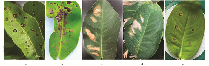
  • 该病害在莲雾果园发病普遍,植株中、下部老叶片受害较重. 发病初期叶片上形成黄色斑纹,或形成一些浅黄白色、圆形或椭圆形的小病斑. 典型病斑呈椭圆形或不规则形,中央区域灰白色,周围有褐色带、黄色带晕圈,中部坏死组织干枯后脱落形成穿孔(图 1abc). 在适合的条件下,叶片上的病斑扩大、联合形成不规则较大病斑或大面积枯死(图 1bde). 发病严重时,病树可见枯枝和落叶,生长发育明显受阻,结果少、果小或不结果. 病菌在果实成熟期侵染,果实表面形成不同大小的圆形、浅褐色至黑褐色病斑,均匀地轮纹状下陷,越是病斑中央下陷越深. 病斑周缘为淡灰黄色带,若遇阴雨天气或空气湿度较高时,病部果肉腐烂,可引起大量落果,病果在运输途中或储藏期间很容易发生腐烂变质.

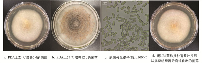
  • 通过分离纯化,从普洱市和西双版纳州采集的病样中各得到3个纯化真菌菌株(分别编码为LWTJ 1,2,3和LB4,5,6),其中分离自普洱市的LWTJ2和来自西双版纳州的LB4基本相同,它们与刺盘孢属真菌的菌落形态近似. 用LWTJ2和LB4离体接种健康莲雾叶片后症状相似,48 h后菌饼下叶片组织呈水浸状坏死,6 d后形成近圆形病斑,其中央呈灰白色,周围黑褐色,褐色坏死部分外围有黄色晕圈,与田间自然发病叶片上典型病斑相似. 在5片叶(重复)上6个接种点全部发病,发病率为100%. 接种10 d后,切取病斑组织10块进行再分离纯化,均成功分离到与原接种菌株菌落形态相同的纯培养物(图 2d). 在相同条件下,其他4个菌株菌落与刺盘孢属真菌菌落有差异,接种后未引起莲雾叶片发病,为污染的杂菌. 因此,根据柯赫氏证病法则确定LWTJ2和LB4菌株为莲雾炭疽病病原菌.

  • LWTJ2和LB4两个致病菌株在PDA培养基上的培养特征基本相同. 在25 ℃恒温箱中培养6 d后,菌落呈圆形,粉白或鹅黄色,边缘光滑齐整,表面呈绒毛状,气生菌丝发达(图 2a);后期(12 d)菌落颜色稍加深,中央区域变成灰黑色,并产生大量蛋黄色分生孢子团(图 2b). 在显微镜下观察,菌丝透明,分隔,呈近直角分枝. 分生孢子无色,呈长椭圆形或柱状,两端盾圆,绝大多数细胞中间部位有一个油球,孢子大小为9.8~18.0(平均13.9±2.1) μm × 4.5~6.0(平均5.5±1.0) μm(图 2c). 在实验室较长期地培养和多次采集莲雾果园自然发病叶片观察,均未发现病菌有性时期的子囊壳及子囊孢子. 病菌的这些形态特征,与Li等[33]描述的暹罗刺盘孢Colletotrichum siamense非常接近.

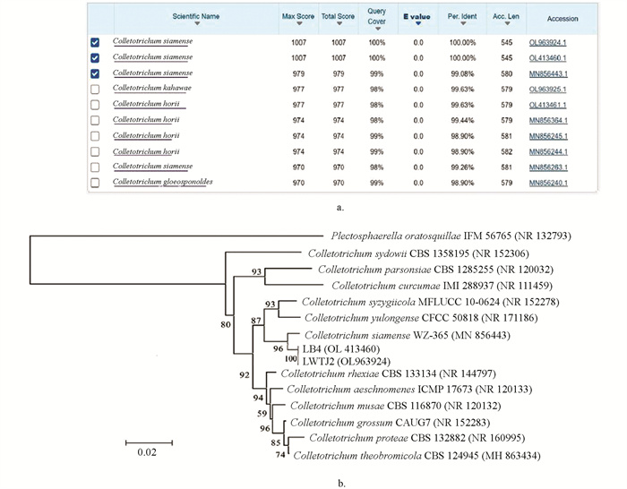
  • 通过PCR扩增获得LWTJ2和LB4两个菌株的基因组rDNA-ITS片段长度均为545 bp,在NCBI GenBank的登录号分别为OL413460和OL963924. Blast-n分析表明这两个菌株ITS序列的最大相似率为100%,它们与C. siamense WZ-365菌株ITS(Acc. No. MN856443)的最大相似率和覆盖率分别为99.08%和99%,与C. kahawae(卡哈瓦刺盘孢)的覆盖率和最大相似率分别为98%和99.63%(图 3a);用MEGA6.0软件[32]绘制LWTJ2和LB4两个菌株ITS与刺盘孢属12种真菌菌株ITS的系统演化树图(图 3b). 图 3b显示,两个菌株与C. siamense WZ-365菌株的ITS(Acc. No. MN856443)聚集在同一末端分枝上;LWTJ2和LB4为同一种真菌(置信度为100%),二者与暹罗刺盘孢为同一物种(置信度为96%). 因此,综合形态特征、ITS序列Blast-n和演化树分析确定,引起莲雾炭疽病的病原菌为暹罗刺盘孢(C. siamense).

3.   讨论与结论
  • 莲雾是一种被广泛栽培的重要果树,目前文献记载的莲雾病害有近60种,其中炭疽病是发生最普遍和危害最严重的一种病害. 莲雾炭疽病是由刺盘孢属Colletotrichum真菌侵染所致,现已报道的有盘长孢刺盘孢C. gloeosporioides、果生刺盘孢C. fructicola和莲雾刺盘C. syzygicola[16-18]. 本研究通过形态学和分子生物学鉴定方法确定,发生在我国云南南部地区的莲雾炭疽病由暹罗刺盘孢C. siamense引起,这与其他国家和地区报道的刺盘孢种类不同,属于莲雾炭疽病的一种新病原真菌.

    暹罗刺盘孢异名有C. melancaulonC. dianeseiC. communisC. endomangiferaeC. hymencallidisC. jasmini-sambac,这些异名来源于不同地区的寄主植物[34-38],均为该真菌的无性型(Anamotph),在早期的分类系统中属于真菌界,半知菌门,腔孢纲,黑盘菌目,黑盘孢科,刺盘孢属. 其有性型(Teleomorph)文献尚无报道,本研究在室内较长时间反复培养及自然发病果树上多次观察寻找均未发现子囊壳和子囊孢子,说明该真菌可能很难产生有性型. 但依据Sutton[39]文献记载,刺盘孢属中很多种类的有性型均属于小丛赤壳属Glomerella,由此推论暹罗刺盘孢的有性型应该是暹罗小丛壳Glomerella siamense,在较新的分类系统中属于真核域、真菌界,双核菌亚界,子囊菌门,子囊菌纲,小丛赤壳目,小丛赤壳科,小丛赤壳属. 此推论将对今后研究或探索暹罗刺盘孢生活史中的有性型阶段和病害循环等具有重要参考作用.

    暹罗刺盘孢是一种寄主范围很广的植物病原真菌,现有文献记载约有60多种炭疽病是由暹罗刺盘孢侵染所致,国外报道的有菠萝蜜、枇杷、无花果、薄荷、胡椒、迷迭香、草莓、辣椒、柑桔、火龙果、芦荟、柿子和美国山核桃等作物的炭疽病[40-45];我国发生的有柑橘、苹果、核桃、番木瓜、荔枝、胡椒、假苹婆、白兰花、鳄梨和越南槐等[46-52]. 本研究鉴定的莲雾炭疽病也由暹罗刺盘孢所致,表明莲雾是暹罗刺盘孢的一个新寄主,在其他莲雾种植国家和地区未曾见报道.

    据课题组2021年的调查结果,莲雾炭疽病在云南省普洱市和西双版纳州各地果园发生普遍,危害较重,明显降低了莲雾果实产量、品质及果农的经济效益. 本研究较详细地观察描述了莲雾炭疽病的症状及其病原的形态特征,鉴定确认了病原菌为暹罗刺盘孢,不仅对该病害田间诊断识别、监测和防控有重要的实用参考价值,而且丰富了莲雾炭疽病病原菌的多样性. 然而,不同植物炭疽病寄主、病原菌及其发生环境条件各有不同,它们的流行规律必然存在较大差异,因此对它们的防控技术措施也会有所不同. 为了减轻或避免莲雾危害和经济损失,促进产业发展,对于莲雾炭疽病流行规律、病原菌生物学特性及其综合防控技术等方面仍有待进一步研究.

    本研究较详细地观察记载了滇南地区莲雾炭疽病症状及其病原菌形态特征,根据形态学与rDNA-ITS序列的Blast-n对比和演化树分析鉴定获得的LWTJ2和LB4两个菌株均属于暹罗刺盘孢C. siamense,这是莲雾炭疽病的一种新病原真菌. 研究结果进一步证明了莲雾炭疽病病原菌的多样性,也为该病害的诊断监测和有效控制提供了科学依据.

Figure (3)  Reference (52)

Catalog

    /

    DownLoad:  Full-Size Img  PowerPoint
    Return
    Return