Message Board

Dear readers, authors and reviewers,you can add a message on this page. We will reply to you as soon as possible!

2022 Volume 47 Issue 2
Article Contents

HUA Jin, ZHENG Ziqiang, YANG Rong, et al. Investigation in Principle on Differential Impacts of Strong-Flavor Liquor and Pure Ethanol on Hepatic Lipid Level[J]. Journal of Southwest China Normal University(Natural Science Edition), 2022, 47(2): 56-62. doi: 10.13718/j.cnki.xsxb.2022.02.011
Citation: HUA Jin, ZHENG Ziqiang, YANG Rong, et al. Investigation in Principle on Differential Impacts of Strong-Flavor Liquor and Pure Ethanol on Hepatic Lipid Level[J]. Journal of Southwest China Normal University(Natural Science Edition), 2022, 47(2): 56-62. doi: 10.13718/j.cnki.xsxb.2022.02.011

Investigation in Principle on Differential Impacts of Strong-Flavor Liquor and Pure Ethanol on Hepatic Lipid Level

More Information
  • Corresponding author: ZHOU Yabin
  • Received Date: 20/02/2020
    Available Online: 20/02/2022
  • MSC: Q26;TS262

  • Liquor is an alcoholic beverage that closely associated with human recreation. However, excessive long-term liquor consumption induces lipid accumulation in liver, and leads to alcoholic liver diseases such as fatty liver disease. In the current study, in order to investigate in principle the potential effects of the volatile flavor substance in liquor on liver lipid levels, the authors utilized an AML12 cell-based liver steatotic model to investigate the effects of three strong-flavor liquor from Sichuan province and ethanol on cellular lipid levels. The volatile components of the three kinds of liquor were also analyzed by HS-SPME-GC-MS and compared using statistical method to analyze the differences in amount. The results show liquor B treatment subgroup (final ethanol concentration of 0.5%) demonstrated significant lower intracellular lipid level than the control group, pure ethanol treatment group (final ethanol concentration of 0.5%) and liquor A treatment subgroup (final ethanol concentration of 0.5%). Statistical analysis of the main flavor substances in liquor A and B show the combining differences in amount of two or more main flavor substances (may also involved main non-volatile substance) may have contributed to the significant difference in cellular lipids levels. In the future studies, it's needed to test the effects on intracellular lipid level of the main flavor substance that are different in amount individually and in combination, and the cellular signalling pathways affected. GC-MS and statistical analysis of the main non-valitile substance is also needed.
  • 加载中
  • [1] 滕抗, 陈杰, 邵海燕, 等. 白酒的起源和造酒学刍议[J]. 酿酒科技, 2013, 229(7): 109-112.

    Google Scholar

    [2] 孙宝国. 国酒[M]. 北京: 化学工业出版社, 2019.

    Google Scholar

    [3] 徐岩, 范文来, 吴群, 等. 风味技术导向白酒酿造基础研究的进展[J]. 酿酒科技, 2012, 211 (1): 17-23.

    Google Scholar

    [4] 王琦. 中国白酒文化与行业发展[J]. 食品工业科技, 2017, 38(5): 14-15.

    Google Scholar

    [5] 徐岩, 范文来, 葛向阳, 等. 科学认识中国白酒中的生物活性成分[J]. 酿酒科技, 2013, 231(9): 1-6.

    Google Scholar

    [6] FANG C, DU H, ZHENG X J, et al. Solid-State Fermented Chinese Alcoholic Beverage (Baijiu) and Ethanol Resulted in Distinct Metabolic and Microbiome Responses[J]. The FASEB Journal, 2019, 33(6): 7274-7288. doi: 10.1096/fj.201802306R

    CrossRef Google Scholar

    [7] 罗强, 刘杰, 刘志刚. 酱香型白酒中活性成分的抗氧化活性[J]. 食品与发酵工业, 2019, 45(23): 84-89.

    Google Scholar

    [8] 黄蕴利, 黄永光, 郭旭. 白酒中的主要生物活性功能成分研究进展[J]. 食品工业科技, 2016, 37(15): 375-379.

    Google Scholar

    [9] YAO H R, LIU J, PLUMERI D, et al. Lipotoxicity in HepG2 Cells Triggered by Free Fatty Acids[J]. American Journal of Translational Research, 2011, 3(3): 284-291.

    Google Scholar

    [10] MORAVCOVÁ A, ČERVINKOVÁ Z, KUČERA O, et al. The Effect of Oleic and Palmitic Acid on Induction of Steatosis and Cytotoxicity on Rat Hepatocytes in Primary Culture[J]. Physiological Research, 2015, 64(suppl 5): 627-636.

    Google Scholar

    [11] LI X L, WANG R, ZHOU N, et al. Quercetin Improves Insulin Resistance and Hepatic Lipid Accumulation in Vitro in a NAFLD Cell Model[J]. Biomedical Reports, 2013, 1(1): 71-76. doi: 10.3892/br.2012.27

    CrossRef Google Scholar

    [12] WU X D, ZHANG L Y, GURLEY E, et al. Prevention of Free Fatty Acid-Induced Hepatic Lipotoxicity by 18β-Glycyrrhetinic Acid through Lysosomal and Mitochondrial Pathways[J]. Hepatology, 2008, 47(6): 1905-1915. doi: 10.1002/hep.22239

    CrossRef Google Scholar

    [13] GLUCHOWSKI N L, BECUWE M, WALTHER T C, et al. Lipid Droplets and Liver Disease: From Basic Biology to Clinical Implications[J]. Nat Rev Gastroenterol Hepatol, 2017, 14(6): 343-355. doi: 10.1038/nrgastro.2017.32

    CrossRef Google Scholar

    [14] TESCHKE R. Alcoholic Liver Disease: Current Mechanistic Aspects with Focus on Their Clinical Relevance[J]. Biomedicines, 2019, 7(3): 68-79. doi: 10.3390/biomedicines7030068

    CrossRef Google Scholar

    [15] BYRNE C D, TARGHER G. NAFLD: a Multisystem Disease[J]. Journal of Hepatology, 2015, 62(1): S47-S64. doi: 10.1016/j.jhep.2014.12.012

    CrossRef Google Scholar

    [16] YOUNOSSI Z, ANSTEE Q M, MARIETTI M, et al. Global Burden of NAFLD and NASH: Trends, Predictions, Risk Factors and Prevention[J]. Nature Reviews Gastroenterology & Hepatology, 2018, 15(1): 11-20.

    Google Scholar

    [17] WANG F S, FAN J G, ZHANG Z, et al. The Global Burden of Liver Disease: The Major Impact of China[J]. Hepatology, 2014, 60(6): 2099-2108. doi: 10.1002/hep.27406

    CrossRef Google Scholar

    [18] 罗欣然, 李秋萍, 舒刚. 保肝口服液对CCl4所致肝损伤小鼠护肝作用评价[J]. 西南师范大学学报(自然科学版), 2021, 46(5): 38-43.

    Google Scholar

  • 加载中
通讯作者: 陈斌, bchen63@163.com
  • 1. 

    沈阳化工大学材料科学与工程学院 沈阳 110142

  1. 本站搜索
  2. 百度学术搜索
  3. 万方数据库搜索
  4. CNKI搜索

Figures(8)  /  Tables(1)

Article Metrics

Article views(1837) PDF downloads(314) Cited by(0)

Access History

Other Articles By Authors

Investigation in Principle on Differential Impacts of Strong-Flavor Liquor and Pure Ethanol on Hepatic Lipid Level

    Corresponding author: ZHOU Yabin

Abstract: Liquor is an alcoholic beverage that closely associated with human recreation. However, excessive long-term liquor consumption induces lipid accumulation in liver, and leads to alcoholic liver diseases such as fatty liver disease. In the current study, in order to investigate in principle the potential effects of the volatile flavor substance in liquor on liver lipid levels, the authors utilized an AML12 cell-based liver steatotic model to investigate the effects of three strong-flavor liquor from Sichuan province and ethanol on cellular lipid levels. The volatile components of the three kinds of liquor were also analyzed by HS-SPME-GC-MS and compared using statistical method to analyze the differences in amount. The results show liquor B treatment subgroup (final ethanol concentration of 0.5%) demonstrated significant lower intracellular lipid level than the control group, pure ethanol treatment group (final ethanol concentration of 0.5%) and liquor A treatment subgroup (final ethanol concentration of 0.5%). Statistical analysis of the main flavor substances in liquor A and B show the combining differences in amount of two or more main flavor substances (may also involved main non-volatile substance) may have contributed to the significant difference in cellular lipids levels. In the future studies, it's needed to test the effects on intracellular lipid level of the main flavor substance that are different in amount individually and in combination, and the cellular signalling pathways affected. GC-MS and statistical analysis of the main non-valitile substance is also needed.

  • 固态法酿造的中国白酒是中国独有的蒸馏酒,拥有2000多年的酿造和饮用历史[1-2]. 白酒是中国传统的发酵食品,不仅蕴含了厚重的中国历史和文化,还富含有众多的风味成分[3-4]. 由于酒精摄入人体后主要通过肝脏进行代谢,肝损伤在饮酒带来的健康问题中占有重要比例,尤其是酒精性脂肪肝. 有研究认为在适度饮用固态法酿造的白酒时,其中含有的多种风味物质可以在一定程度上降低酒精对人体的伤害,因此其对人体健康的影响与单纯的酒精不同[5-6]. 目前关于白酒对健康影响的研究文献多着眼于分析白酒风味物质中的活性成分,通过活性成分本身的功能推测其在白酒中可能起到的对健康的影响[7-8]. 本研究使用基于小鼠肝实质细胞(AML12)的肝脂质聚集模型[9],对比研究几种浓香型川酒与单纯酒精对肝脂质代谢的影响,检测分析这几种川酒的主要风味物质,并对主要挥发性成分的差异用统计学方法进行分析,研判其是否具有显著性,结果将为白酒中的风味物质对肝脏脂质水平的影响提供数据和研究模型.

1.   材料与方法
  • 四川某品牌浓香型白酒A(酒精浓度为52%),四川某品牌浓香型白酒B(酒精浓度为52%),四川某品牌浓香型白酒C(酒精浓度为52%).

    所用试剂及药品购于美国Sigma Aldrich公司,实验用水皆为去离子水.

    气相色谱质谱联用仪(GC-MS联用仪6890N-5975B inert XL EI/CI MSD),美国Agilent公司; 正倒置一体显微镜(Revolve G176),美国Echo公司.

  • 准确量取900 μL酒样,并加入100 μL 2-辛醇内标溶液,将顶空进样瓶密封,直接自动进样.

  • Agilent J&W DB-WAX气相色谱柱(122-7062),柱长60 m,内径250 μm,膜厚度0.25 μm. 升温程序: 起始温度38 ℃,以1 ℃/min升温至60 ℃,保持2 min; 以3 ℃/min升温至70 ℃,保持2 min; 以4 ℃/min升温至105 ℃,保持2 min; 以4 ℃/min升温至180 ℃,保持1 min; 以6 ℃/min升温至230 ℃,保持2 min. 汽化室温度250 ℃; 载气He,柱前压15.92 psi,载气流量1 mL/min,进样量1 μL; 不分流进样.

  • 质谱的电离方式为电子轰击离子源(EI). 离子源温度230 ℃,四极杆温度150 ℃,电子能量70 eV,发射电流34.6 μA,倍增器电压1 470 V,进样口温度230 ℃,扫描质量范围20~550 amu,溶剂延迟3 min.

  • 用血球计数板法测定小鼠正常肝细胞(AML12)浓度,取35 mm培养皿,每个培养皿里接种1× 10⁵个肝细胞,放置培养箱中(37 ℃,5% CO2)培养24 h. 然后将肝脏细胞样品分别进行如下处理: ①脂质处理组,只用棕榈酸和油酸处理细胞; ②四川浓香型白酒处理组: 分为3个不同白酒处理组,在脂质处理的基础上分别加入3种不同品牌的浓香型白酒A,B和C. 每个白酒处理组又分为两个不同剂量亚组,即在培养液中最终酒精浓度为1%和0.5%; ③酒精处理组: 该组在脂质处理的基础上添加纯酒精并分为两个剂量亚组,分别是在培养液中最终酒精浓度为1%和0.5%; ④无处理空白对照组: 该组不进行脂质、添白酒和纯酒精处理. 细胞进行各种处理后在培养箱中(37 ℃,5% CO2)培养24 h,然后采用尼罗红(染色细胞脂滴中的中性脂)和Hoechst33342(染色细胞核)对细胞内的脂滴和细胞核同时避光进行双染色20 min,用Revolve G176正倒置一体显微镜和ImagJ软件使用预先设定的方法测定肝脏细胞内被尼罗红染色脂滴的荧光强度和Hoechst33342染色的肝脏细胞核数,并计算每个肝脏细胞的平均脂滴荧光强度.

  • 脂质处理组、各浓香型白酒处理(亚)组、酒精对照(亚)组、空白对照组均设置3个平行样品,实验进行3次重复,取3次重复实验平均值(x±s)作为实验结果. 白酒酒样的检测进行3次独立实验,取3次实验平均值(x±s)作为实验结果. 对比各处理组之间细胞脂质水平差异使用One way ANOVA,各处理组1%和0.5%亚组之间细胞脂质水平差异使用双尾Students’T test,白酒A和B之间风味物质差异使用双尾Students’T test进行统计学分析.

2.   结果与分析
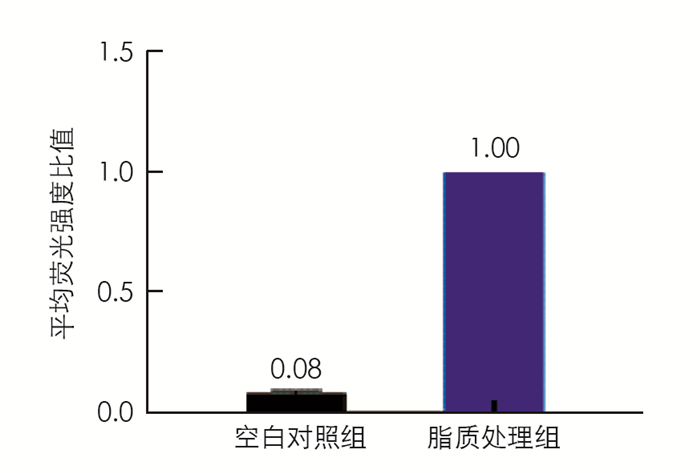
  • 本研究基于油酸和棕榈酸处理肝细胞造成细胞脂质聚集模型[10]检测不同药物处理对肝细胞内脂质的聚集水平,实验结果显示脂质处理组的细胞中脂质水平是空白对照组细胞的12.5倍,表明细胞中形成了脂质聚集(图 1).

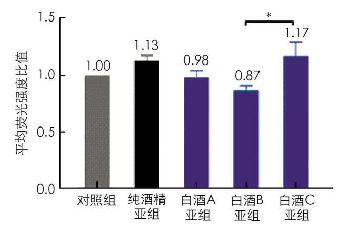
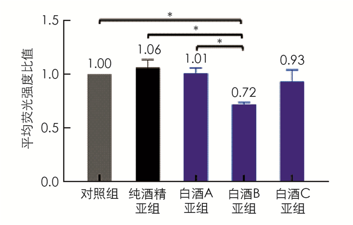
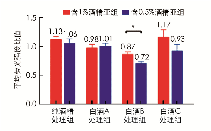
    对脂质聚集的细胞分别用酒精或3种不同浓香型川酒进行处理,含1%酒精处理亚组中,白酒B亚组的脂质水平为0.87,显著低于白酒C亚组的脂质水平(1.17,p < 0.05); 脂质处理组(以下简称对照组)的脂质水平要低于纯酒精亚组,白酒A亚组和白酒B亚组细胞内脂质也低于纯酒精亚组,但差异无统计学意义(图 2). 含0.5%酒精的处理亚组中白酒B亚组的脂质水平(0.72)显著低于对照组(p < 0.05)、纯酒精亚组(p < 0.05)和白酒A亚组(p < 0.05),其他各组的脂质水平差异无统计学意义(图 3). 同种处理的亚组中,除白酒A外,含0.5%酒精的亚组的脂质水平均要低于含1%酒精的亚组(图 4),其中白酒B含0.5%酒精亚组的脂质水平显著低于含1%酒精亚组的脂质水平(p < 0.05).

  • 通过GC-MS分别从3种浓香型白酒中检测出的风味物质较丰富,有33种醇类(图 5)、27种酯类(图 6)、18种醛类、酮类和酸类(图 7)以及20种其他物质如乙缩醛等(图 8). 3种白酒虽然同是浓香型白酒,但在醇、酯、酸和醛等风味物质的种类和相对浓度上还是有一定的差异.

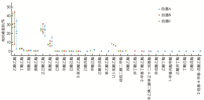
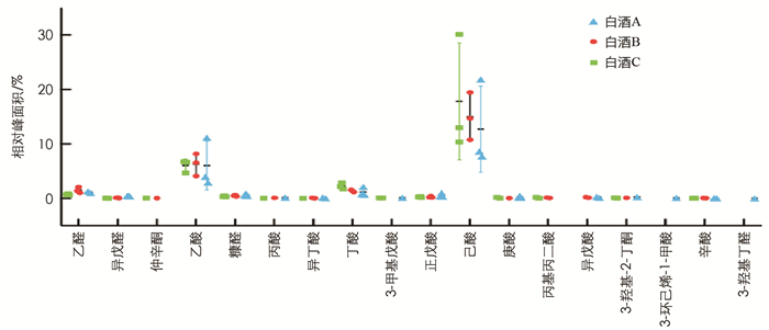
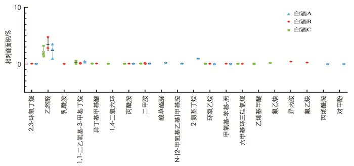
    首先,含0.5%酒精的白酒B亚组中脂质水平显著低于白酒A亚组(p < 0.05),而与白酒C亚组差异无统计学意义,因此对白酒A和B中均含有的挥发性风味物质(相对浓度1%或以上)进行分析比较,结果显示其相对浓度有一定的差异(表 1). 如在醇类物质中,白酒A中检测到明显高于白酒B的丙醇(分别为2.57%和0.73%,p < 0.001)、正丁醇(分别为3.02%和1.00%,p < 0.001)和异戊醇(分别为3.04%和0.99%,p < 0.001). 两种白酒都含有较高的乙酸乙酯、正己酸乙酯和乳酸乙酯,但差异均无统计学意义. 其中正己酸乙酯在两种白酒中的相对浓度差异不大,而白酒B中的乙酸乙酯(27.52%)要高于白酒A(20.14%),白酒B中乳酸乙酯(8.65%)要低于白酒A(11.16%). 乙酸和己酸在两种白酒的挥发性风味物质中的比重也较高,但差异无统计学意义. 乙缩醛在白酒B中的相对浓度(3.47%)要高于白酒A(2.38%),但差异无统计学意义.

    此外,3种白酒均含有其特有的,即其他两种白酒中未检测到的挥发性风味物质. 如白酒C含有4.46%的2-辛醇,而在白酒A和B中未检出; 白酒A含有在白酒B和C中未检出的己酸丙酯(0.07%); 异丁酸乙酯只在白酒B中有检出(0.18%).

    还有个别风味物质如4-甲基-1-戊醇只在白酒A与B中有检出,甲酸己酯只在白酒A与C中有检出,而仲辛酮只在白酒B和C中有检出.

3.   讨论
  • 本研究发现两个不同剂量的纯酒精处理亚组和白酒A与C亚组的细胞脂质水平与对照组差异无统计学意义. 白酒B(含0.5%酒精)亚组的细胞脂质水平是各组中最低的,且显著小于对照组(p < 0.05)和0.5%纯酒精亚组(p < 0.05),这很有可能是由白酒B中非酒精组分造成的; 白酒B(含0.5%酒精)亚组的细胞脂质水平显著低于白酒A(含0.5%酒精)亚组(p < 0.05),进一步表明两组脂质水平的差异可能是由白酒B与白酒A中有差异的非酒精成分造成的. 对比白酒A与白酒B中主要挥发性风味物质发现,差异有统计学意义的风味物质相对浓度在白酒A中均高于白酒B. 而相对浓度超过5%的乙酸乙酯、正己酸乙酯、乳酸乙酯、乙酸和己酸(乳酸乙酯除外)在白酒B中相对浓度均高于白酒A,但差异无统计学意义. 这表明白酒B与白酒A对细胞脂质水平影响的差异可能不是由某一种相对浓度有差异的风味物质造成,而可能是由两种或以上的风味物质共同作用造成,其中也有可能包含有相对浓度较高的非挥发性物质.

    纯酒精、白酒B和白酒C处理组中均呈现出随酒精相对浓度从1%减低到0.5%其脂质水平降低的趋势,尤其是白酒B,脂质水平降低有统计学意义(p < 0.05),这表明,酒精本身对细胞脂质水平存在影响.

    本研究使用的模型是脂肪肝的细胞模型[10-12],脂质代谢是肝脏的重要功能之一[13]. 肝脏脂质代谢紊乱会引起脂肪肝,脂肪肝的病理起源除了长期酗酒外[14],还有一类是由于生活和饮食习惯引起的非酒精性脂肪肝[15-16],中国目前有近30%的人群是非酒精性脂肪肝患者[17],因此研究饮酒对非酒精性脂肪肝的影响及其机理也是十分必要的.

4.   结论
  • 肝细胞的脂质代谢和其脂质水平与人体健康息息相关[18]. 本研究使用基于AML12肝细胞的脂质聚集模型初步探究了3种不同浓香型白酒对肝细胞内脂质水平的影响,结果显示白酒B对肝细胞脂质水平的降低可能是由两种或以上主要挥发性风味物质组合(可能也包括浓度较高的非挥发性物质)造成. 接下来的研究一方面进一步测试主要风味物质单独和组合对肝细胞脂质水平的影响,探究其影响的细胞代谢路径; 另一方面使用GC-MS联用检测并比较非挥发性风味物质.

Figure (8)  Table (1) Reference (18)

Catalog

    /

    DownLoad:  Full-Size Img  PowerPoint
    Return
    Return